

Hulya Ozturk ( Department of Pediatric Surgery, Turkey. )
Hayrettin Ozturk ( Department of Pediatric Surgery, Turkey. )
Elcin Hakan Terzi ( Department of Histology and Embryology, Turkey. )
Arif Duran ( Department of Emergency, Abant Izzet Baysal University, Medical School, Bolu, Turkey. )
Ibrahim Uygun ( Department of Pediatric Surgery, Dicle University, Medical School, Diyarbakir, Turkey. )
Ufuk Ozgen ( Department of Pharmacognosy, Faculty of Pharmacy, Ataturk University, Erzurum, Turkey. )
March 2014, Volume 64, Issue 3
Original Article

Abstract
Objective: To investigate the potential protective effects of Rosmarinic acid (RA) on rats exposed to ischaemia/reperfusion renal injury.
Methods: The prospective study was conducted at Abant Izzet Baysal University, Turkey, and comprised 21 male Spraque Dawley rats weighing 250-270g each. They were divided into three equal groups. Unilaterally nephrectomised rats were subjected to 60 minutes of left renal ischaemia followed by 60 minutes of reperfusion. Group 1 had sham-operated animals; group 2 had ischaemia/reperfusion untreated animals; and group 3 had ischaemia/reperfusion animals treated with rosmarinic acid. Serum creatinine, blood urea nitrogen, tissue malondialdehyde, glutathione peroxidase, superoxide dismutase and myeloperoxidase (MPO) activities, and light microscopic findings were evaluated. SPSS 17 was used for statistical analysis.
Results: Treatment of rats with rosmarinic acid produced a reduction in the serum levels of creatinine and blood urea nitrogen compared to the other groups. However, no statistically significant difference was found. The levels of malondialdehyde and myeloperoxidase were decreased in the renal tissue of group 3, while glutathione peroxidose and superoxide dismutase levels remained unchanged. The injury score decreased in the treatment group rats compared to the untreated group. Rosmarinic acid significantly decreased focal glomerular necrosis, dilatation of Bowman\'s capsule, degeneration of tubular epithelium, necrosis in tubular epithelium, and tubular dilatation.
Conclusions: Rosmarinic acid prevented ischaemia/reperfusion injury in the kidneys by decreasing oxidative stress.
Keywords: Rat, Renal/kidney, Ischaemia/Reperfusion injury, Rosmarinic acid. (JPMA 64: 260; 2014).
Introduction
Acute renal failure occurring in cases such as transplantation, shock, sepsis, and renal artery stenosis is a major clinical problem. Therefore, it is usually associated with high morbidity and mortality.1,2
Ischaemia-reperfusion (I-R) injury leads to cell damage, cell death, increased vascular permeability, tissue necrosis, and multiorgan dysfunction. These pathophysiological processes include activation of neutrophils, platelets, cytokines, reactive nitrogen species, reactive oxygen substances (ROS), the coagulation system, the endothelium, and the xanthine-oxidoreductase enzyme system. Cell death as a result of both necrosis and apoptosis is triggered by substances released during I-R injury.3-5
Several natural products have been reported to have beneficial effects on I-R injury, particularly from a preventative perspective. Rosmarinic acid (RA), a phenolic compound found in various Labiatae herbs,5 possesses anti-inflammatory and anti-oxidative properties.6-8 However, no study has ever been performed on kidney tissue. In the present study, we investigated the protective effect of RA on oxidative stress, renal dysfunction, and histologic alterations induced by renal I-R in rats.
Material and Methods
Approved by the Animal Care Committee of Abant Izzet Baysal University, Bolu, Turkey, the study was performed in accordance with National Institute of Health guidelines for the use of experimental animals.
The study used 21 male Spraque Dawley rats weighing 250-270g each. They were housed individually in a room maintained at a constant temperature (24±2°C) and humidity (55±15%) and were randomly assigned to 3 equal groups. The rats were allowed free access to standard rat chow and water before and after the experiment. The animals were kept on a fast overnight before the experiments, but were given free access to water. They were anaesthetised by 100-mg/kg ketamine and 20-mg/kg xylazine body weight, i.p. Right femoral vein was cannulated for administration of drugs and saline.
In the sham-control group, the abdomen was dissected, and the right kidney was harvested, and then the left renal pedicle was exposed. Renal clamps were not applied. In the I-R group, the right kidney was harvested and then the left renal artery and vein were clamped with a homeostasis clip for 60 minutes. The abdomen was closed during I-R. The clip was subsequently removed to permit reperfusion for 60 minutes. In the I-R/RA treated group, the same surgical procedure as in group II was performed. RA 25mg/kg, was intraperitoneally administered 30 minute prior to the induction of ischaemia.
Blood samples (4-5ml) were collected at 60 minutes after reperfusion and the rats were sacrifised afterwords while the kidney tissue was harvested.
Blood urea nitrogen (BUN) and creatinine were measured as indicators of disorders of glomeruli and endothelial cell. The activities of BUN and creatinine in plasma were determined by standard auto-analyser methods on an Abbot Aeroset (USA).
Biochemically, malondialdehyde (MDA) contents of the homogenates were determined spectrophotometrically by measuring the presence of thiobarbituric acid reactive substances.9 Three ml of 1% phosphoric acid and 1 ml 0.6% thiobarbituric acid solution were added to 0.5 ml of homogenate pipetted into a tube. The mixture was heated in boiling water for 45 min. After the mixture cooled, the coloured part was extracted into 4 ml of n-butanol. The absorbance was measured by spectrophotometer (UV-1601; Shimadzu, Kyoto, Japan) at 532 and 520 nm. The amount of lipid peroxides was calculated as thiobarbituric acid reactive substances of lipid peroxidation. The results were expressed in nmol/g tissue, according to a prepared standard graph. Total superoxide dismutase (SOD) activity was determined according to the method of Sun et al. 10 The principle of the method is the inhibition of nitroblue tetrazolium (NBT) reduction by the xanthine-xanthine oxidase system as a superoxide generator. One unit of SOD was defined as the amount of enzyme causing 50% inhibition in the NBT reduction rate. SOD activity was expressed in U/g protein. Glutathione peroxidase (GPx) activity was measured by the method of Paglia and Valentine.11 An enzymatic reaction in a tube containing Nicotinamide adenine dinudcotide phosphate (NADPH), reduced glutathione (GSH), sodium aside and glutathione reductase was initiated by adding H2O2; the change in absorbance at 340 nm was observed using a spectrophotometer. The activity was expressed as U/mg protein. The homogenates were then centrifuged at 17,000 x g at 4°C for 15 min, and myeloperoxidase (MPO) activity (U/g protein) in the supernatant was measured as described in literature.12
For histopathological evaluation, one of the pieces were fixed in 10% buffered formalin, and embedded in paraffin. Sections were cut at 5µm and coded kidney specimens were stained with haematoxylin and eosin and examined in a blinded fashion. Histological changes were evaluated by quantitative measurement of tubulointerstitial injury, which was assessed by counting the number of necrotic and apoptotic cells, loss of tubular brush border, tubular dilatation, cast formation, and neutrophil infiltration. The scoring was 0 = none; 1=10%; 2= 11% to 25%; 3 = 26% to 45%; 4 = 46% to 75%; and 5 =76%.12
All statistical analyses were carried out using SPSS version 17.0. All data was presented as mean ± standard deviation. Differences in measured parametres among the three groups were analysed by the Kruskal-Wallis test. Dual comparisons between groups that presented significant values were evaluated with the Mann-Whitney U-test. Statistical significance was accepted at a value of p<0.05.
Results
The mean values of BUN were 19±1.9, 27±2.8, 22±2.1 mg/dL in the three groups, respectively. Likewise, the mean values of creatinine were 0.3±0.1, 1.5±0.3, 1.1±0.2 mg/dL in the three groups. In rats treated with RA, a reduction in the serum levels of creatinine and BUN was determined compared to the untreated group, but the difference was not statistically significant.
The values of oxidative stress-associated parametres were noted (Figure-1).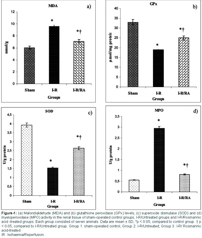
The values of MDA and MPO were decreased in the I-R/RA group (p < 0.001) compared to the I-R group. Also, in the RA-treated group, GPx and SOD levels were found to be unchanged (p < 0.05).
The renal injury score increased in experimental groups compared to the sham-control (p<0.001, p<0.001, respectively). In the RA treatment group, the renal injury score decreased compared to the I-R group (p<0.01) (Figure-2).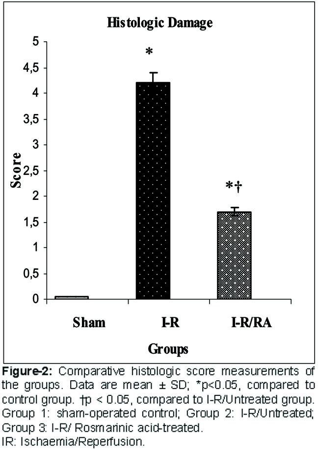
In the sham-control group, there were normative histological changes (Figure 3A).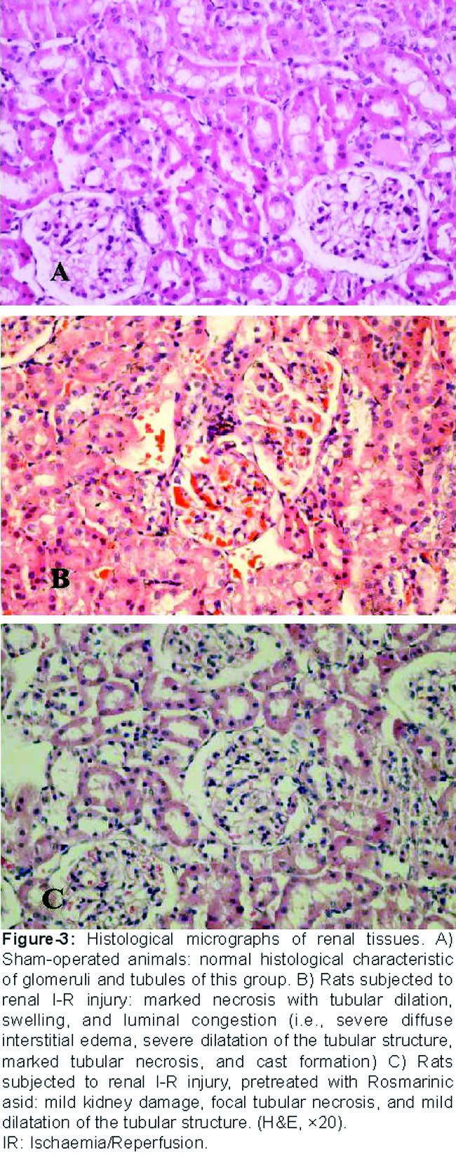
In the I-R group, focal glomerular necrosis, dilatation of Bowman\'s capsule, degeneration of tubular epithelium, necrosis in tubular epithelium, and tubular dilatation were found (Figure 3B). In the RA treated group, decreased focal glomerular necrosis, dilatation of Bowman\'s capsule, degeneration of tubular epithelium, and necrosis in tubular epithelium were noticed (Figure-3C).
Discussion
I-R injury of the kidney causes severe tissue damage, with typical functional and pathophysiological characteristics, including diminished glomerular filtration rate, tubular sodium and potassium re-absorption and renal blood flow.13,14 In the present study, we demonstrated that RA reduces both the glomerular and the tubular injury and dysfunction caused by severe ischaemia in rats assessed by histopathological and biochemical inflammatory parametres.
Reperfusion of the kidney after ischaemia causes the release of pro-inflammatory substances and the formation of both reactive nitrogen species and ROS, free radicals, such as superoxide, peroxide, and hydroxyl radicals.14 These pro-inflammatory substances and free radicals are normally removed by antioxidant enzymes, such as SOD and GPx. MDA, frequently used to show the involvement of free radicals in cell damage, is one of the final products of lipid peroxidation.15,16 RA, a phenolic compound found in various Labiatae herbs,6 possesses several anti-inflammatory properties and anti-oxidant activities.17,18 It was isolated from many species of the families Lamiaceae and Boraginaceae and was identified as one of the active components of several medicinal plants (e.g. Salvia officinalis, Mentha x piperita, Thymus vulgaris, Melissa officinalis, Symphytum officinale) within these families.19 The anti-oxidative property of RA has been demonstrated by its ability to reduce liver injury induced by D-galactosamine and lipopolysaccharides through the scavenging of superoxide molecules and the inhibition of cyclo-oxygenase-2.7,8,20 Pérez-Fons et al21 suggested that hydrophobic diterpenes and the hydrophilic RA were responsible for the anti-oxidant capacity of rosemary leaf extracts. Makino et al22 found that RA exhibited anti-proliferative effects in cultured murine mesangial cells and suppressive effects on platelet-derived growth factor (PDGF) and c-myc mRNA expression. C-myc mRNA is a type of mRNA that serves as a template for the MYC proten which is implicated in the rapid growth of cancer cells. It is tempting to speculate that RA in Labiatae herbs is a promising agent to prevent mesengioproliferative disease.
The present study found that ROS enzyme activity decreased after I-R injury, suggesting that the free radical scavenging system was destroyed by I-/R process. In contrast, tissue anti-oxidant enzymes, SOD, and GPx were significantly higher in the rats given RA. These results suggest that the levels of various anti-oxidant enzyme (serum SOD, GPx) levels, which protect against oxygen-free radicals, were higher in the RA-treated group. Generally, the conversion of the superoxide anion and hydrogen peroxide was impaired due to the decreased levels of SOD, and GPx, resulting in an increase in the level of oxygen-free radicals. Therefore, the elevated superoxide and hydrogen peroxide levels accelerate the damage. In this study, RA as an exogenous anti-oxidant appeared to attenuate I-R injury by increasing the activities of serum SOD and GPx. The histologic examination showed less damage to the tubules in the RA treatment group than in the I-R group. It has been reported that an inflammatory response induced by ischaemia followed by reperfusion is largely responsible for the tissue damage observed.14-16 In our study, treatment with RA prior to the I-R process had better morphological features. Our study had some limitations. For example, we studied the therapeutic effects of RA for only 1h after reperfusion. It is possible that the pharmacological effects of these compounds would have been different at time points further out from the reperfusion event. Additionally, in this preliminary investigation, we studied only one dosage amount of the compound, and it is quite possible that the dose-response relationships for this compound may differ substantially.
Conclusion
The current study shows for the first time that RA prevented I-R injury in the kidneys by decreasing oxidative stress. The administration of RA before renal ischaemia may decrease the severity of I-R injury by its anti-oxidant capacity and free radical scavenging activity. Clinical trials are needed to reveal further benefits of this natural anti-oxidant in the renal I-R injury.
References
1. Rondon-Berrios H, Palevsky PM. Treatment of acute kidney injury: An update on the management of renal replacement therapy. Curr Opin Nephrol Hypertens 2007; 16: 64-70.
2. Yazihan N, Ataoglu H, Kavas GO, Akyurek N, Yener B, Aydm C. The effect of K ATP channel blockage during erythropoietin treatment in renal ischemia-reperfusion injury. J Invest Surg 2008; 21: 340-7.
3. Fan LH, He L, Cao ZQ, Xiang B, Liu L. Effect of ischemia preconditioning on renal ischemia/reperfusion injury in rats. Int Braz J Urol 2012; 38: 842-54.
4. Sedaghat Z, Kadkhodaee M, Seifi B, Salehi E, Najafi A, Dargahi L. Remote preconditioning reduces oxidative stress, downregulates cyclo-oxygenase-2 expression and attenuates ischaemia-reperfusion-induced acute kidney injury. Clin Exp Pharmacol Physiol 2013; 40: 97-103.
5. Salehipour M, Monabbati A, Salahi H, Nikeghbalian S, Bahador A, Marvasti VE, et al. Protective effect of parenteral vitamin E on ischemia-reperfusion injury of rabbit kidney. Urology 2010; 75: 858-61.
6. Makino I, H Tanaka. From a choleretic to an immunomodulator: historical review of ursodeoxycholic acid as a medicament. J Gastroenterol Hepatol 1998; 13: 659-64.
7. Psotova J, Kolar M, Sousek J, Svagera Z, Vicar J, Ulrichova J. Biological activities of Prunella vulgaris extract. Phytother Res 2003; 17: 1082-7.
8. van Kessel KP, E Kalter S, Verhoef J. Rosmarinic acid inhibits external oxidative effects of human polymorphonuclear granulocytes. Agents Actions 1986; 7: 375-6.
9. Uchiyama M, Mihara M. Determination of malonaldehyde precursor in tissues by tiobarbituric acid test. Anal Biochem 1978; 34: 271-8.
10. Sun Y, Oberley L, Li Y. A simple method for clinical assay of superoxide dismutase. Clin Chem 1988; 34: 497- 500.
11. Paglia DE, Valentine WN. Studies on the quantitative and qualitative characterization of erythrocyte glutathione peroxidase. J Lab Clin Med 1967; 70: 158-70.
12. Schierwagen C, Bylund-Fellenius AC, Lundberg C. Improved method for quantification of tissue PMN accumulation measured by myeloperoxidase activity. J Pharmacol Meth 1990; 23: 179-86.
13. Feng L, Xiong Y, Cheng F, Zhang L, Li S, Li Y. Effect of ligustrazine on ischemia-reperfusion injury in murine kidney. Transplant Proc 2004; 36: 1949-51.
14. Chatterjee PK. Novel pharmacological approaches to the treatment of renal ischemia-reperfusion injury: A comprehensive review. Naunyn Schmiedebergs Arch Pharmacol 2007; 376: 1-43.
15. Erdogan H, Fadillioglu E, Yagmurca M, Ucar M, Irmak MK. Protein oxidation and lipid peroxidation after renal ischemiareperfusion injury: Protective effects of erdosteine and N-acetylcysteine. Urol Res 2006; 34: 41-6.
16. Li C, Jackson RM. Reactive species mechanisms of cellular hypoxia-reoxygenation injury. Am J Physiol Cell Physiol. 2002; 282: C227-C41.
17. Peake PW, Pussell BA, Martyn P, Timmermans V, Charlesworth JA. The inhibitory effect of rosmarinic acid on complement involves the C5 convertase. Int J Immunopharmacol 1991; 13: 853-7.
18. Qiao S, Li W, Tsubouchi R, Haneda M, Murakami K, Takeuchi F, et al. Rosmarinic acid inhibits the formation of reactive oxygen and nitrogen species in RAW264.7 macrophages. Free Radical Res 2005; 39: 995-1003.
19. Petersen M, Abdullah Y, Benner J, Eberle D, Gehlen K, Hücherig S, et al. Evolution of rosmarinic acid biosynthesis. Phytochemistry 2009; 70: 1663-79.
20. Won J, Hur YG, Hur EM, Park SH, Kang MA, Choi Y, et al. Rosmarinic acid inhibits TCR-induced T cell activation and proliferation in an Lck-dependent manner. Eur J Immunol 2003; 33: 870-9.
21. Pérez-Fons L, Garzón MT, Micol V. Relationship between the antioxidant capacity and effect of rosemary (Rosmarinus officinalis L.) polyphenols on membrane phospholipid order. J Agric Food Chem 2010; 58: 161-71
22. Makino T, Ono T, Muso E, Yoshida H, Honda G, Sasayama S. Inhibitory effects of rosmarinic acid on the proliferation of cultured murine mesangial cells. Nephrol Dial Transplant 2000; 15: 1140-5.
Related Articles
Journal of the Pakistan Medical Association has agreed to receive and publish manuscripts in accordance with the principles of the following committees:


