

Mohammad S. Humayun ( Department of Surgery, Al-Jahra Hospital, Kuwait. )
Aly M. Rady ( Department of Surgery, Al-Jahra Hospital, Kuwait. )
June 1987, Volume 37, Issue 6
Case Reports

CASE REPORT
A 40 year old lady with 3 children was admitted with a 2 year history of vague pain in the epigastrium without radiation, relation to meals or jaundice. Occasionally it was associated with vomiting and fat intolerance. Menstrual periods were regular and she had never been on contraceptive tablets or other hormones. She was neither diabetic nor hypertensive.
On examination, the B.P. was 120/70 mm. Hg. and pulse 72/mi Abdominal examination revealed a non-tender hepatic mass measuring 12 cm x 12 cm, enlarged spleen and a small paraumbilical hernia. The Hb level was 9.6 G/dl, W.B.C. 3,500 per c.cm with a normal differential and piatsiet count and coagulation profile. Scanning for sickling and red cell fragility were normal as were serum electrolytes, urea, liver enzymes and total proteins. Serum bilirubin was 1.6 mg/dl and serum hon 10 mgs/dl (42-1 35) but no cause for iron loss was detected. Indirect haemagglutination test for hydatid, alphafeto protein and 5 H.I.A.A., were also normal.
Plain X-ray of the abdomen showed an enlarged spleen with calcification in the splenic and mesenteric arteries and a soft tissue mass in the liver region. Barium enema was normal but a barium meal examination revealed medial displacement of the duodenum. Intravenous pyelography showed that the middle third of the right ureter was dilated, irregular and displaced laterally. The urinary bladder showed a smooth extrinsic impression on the right side. Ultrasound examination showed enlargement of the liver and spleen with an echogenic mass related to the right lobe of the liver, liver scan showed hepatosplenomegaly with a large focal defect in the right lobe. Midstream aortography revealed a dilated tortuous splenic artery with massive splenic enlargement, upward displacement of the hepatic artery and corkscrew appearance of its terminal branches. This displacement was due to a large smooth mass which showed no abnormal vascularity. The venous phase showed tortuous dilated splenic veins. Multiple calcified shadows were seen in the portal venous radicles in the right iliac fossa and another retroperitoneal mass displaced the right ureter laterally (Figure 1).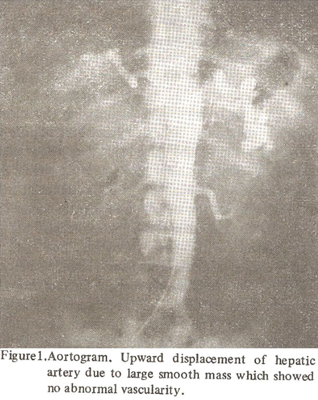
Laparotomy performed on 309.83 revealed a smooth uniform well-circumscribed and encapsulated mass of 12 cm x 12cm in the right lobe of the liver on its anterio-inferior surface with long base and micronodular type cirrhosis of the liver (Figure 2).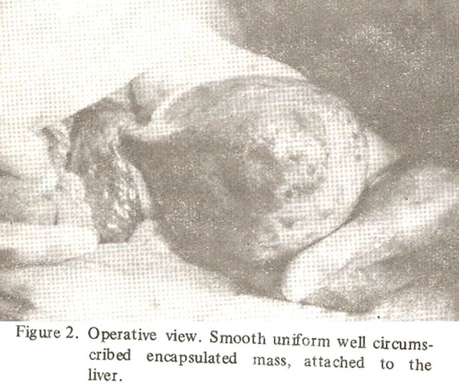
The splenomegaly was confirmed together with plexiform retroperitoneal variceal veins in the right iliac fossa with calcification in their walls. The removed appendix was small 1.5cm, greyish fibrosed and granular.
Histopathological examination described the liver mass as “Globoid, weighs 640 gms and measures 13 x 10 x 7cm. It had a narrow pedicle of 1.5cm and was encapsulated except at its pedicle. Inside this capsule, there was a zone of compressed and nodular liver tissue 0.5 -1 cm in thickness completely surrounding the tumour nodule measuring 10 x 8 x 6cm. This nodule was distinctly delineated from the liver tissue on all sides by either a thin fibrous capsule or merely a difference of appearance and architecture. The consistency of the nodule was slightly softer than the liver. The colour was greyish and sharply contrasted with the yellowish hue of the surrounding rim of the liver tissue.” Microscopically, the tumour was sharply distinct from the surrounding liver and a fibrous capsule was only seen in part. The tumour consisted uniformly of anastomosing cords of liver cells, supported by a reticulin framework. Thin walled blood vessels were seen but no portal tracts. There was no atypia or vascular invasion. By all the known criteria, the tumour was an adenoma of the liver. The rim of the liver tissue showed the changes of cirrhosis’ (Figure 3a).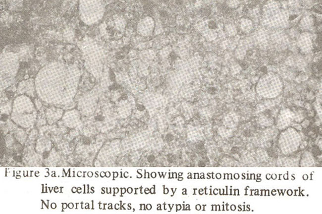
The entire 1.5cm length of the appendix was replaced by carcinoid tumour. Argyrophil and argentaffIn granules were seen (Figure 3b).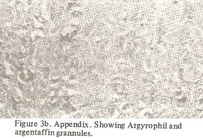
DISCUSSION
Patients with benign hepatomas can present with a variety of clinical features. A mass or pain in nght upper quadrant of symptoms of acute intraperitoneal haemorrhage are common presentations.5-7 Routine radio logical and laboratory studies may not be helpful. Liver scan may demonstrate a focal defect but cannot distinguish between benign and malignant neoplasm unless hepatic arteriography is also performed. Dilated hepatic arteries with irregular tumour vessels, vascular lakes and arteriovenous shunting are suggestive of malignancy and hypovascular tumours show only arterial displacement or encasement. The angiographic findings of benign hepatomas are a clearly defined margin and a peripheral arterial supply with approximately parallel centrally penetrating arteries.8
REFERENCES
1. Edmondson, H.A., Henderson, B. and Benton, B. Liver-Cell adenomas associated with use of oral contraceptives. N. Engi. J. Med., 1976;294:470.
2. Tountas, C., Paraskevas, G. and Deligeorgi, H. Benign hepatomas and oral contraceptives. Lancet, 1974;1 : 1351.
3. Vosnides, G., Brander, W., O’Keeffe, B., Bewick, M. and Ogg, C. Liver hamartomas in patients on oral ‘contraceptives. Br. Med. J 1974; 3:580.
4. Andersen, P.H. and Packer, J.T. Hepatic adenoma; observation after oestrogen withdrawal. Arch. Surg., 1976; 111:898.
5. Syracuse, D.C., Perzin, K.H., Price, J.B., Wiedel, P.D. and Mesa-Tejada, R. Carcinoid tumour of the appendix Mesoappendiceal extension and nodal metastases. Ann. Surg., 1979; 190 :58.
6. Henson, S.W. Jr., Gray, H.K. and Dockerty, MB. Benign tumours of the liver. I. Adenomas. Surg. Gynecol. Obstet., 1956; 103:23.
7. Baum, J.K., Holtz, F., Bookstein, J.J. and Klein, E.W. Possible association between benign hepa. tomas and oral contraceptives. Lancet, 1973; 2: 926.
8. Franklin, R.G. and Downing, C.F. Primary liver tumours. Am. J. Surg., 1947; 7 3:390.
Journal of the Pakistan Medical Association has agreed to receive and publish manuscripts in accordance with the principles of the following committees:


