

Mahfooz Akhtar ( Atomic Energy Medical Centre, Nishtar Medical College and Hospital, Multan, Pakistan. )
Durr-e-Sabih ( Atomic Energy Medical Centre, Nishtar Medical College and Hospital, Multan, Pakistan. )
November 1989, Volume 39, Issue 11
Case Reports

Ultrasound is extensively used in our Centre to image abdominal viscera and cardiac structures. We also use sonography to visualize pleural lesions1. This report describes the unusual appearance of an epidermoid carcinoma of the lung as seen on sonography.
CASE REPORT
A 60 years old male presented with chest pain localized to the right upper chest. On investigations, his ESR was 100 mm, leucocyte count of 18500/mm3 with a polymorphonuclear cell count of 85%. An X-ray chest showed a well defined lesion in upper and mid zone of the right lung (Figure 1).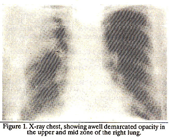
Lung scan showed reduced perfusion of the same region. Ultrasound showed a rounded, well defined relatively an-echoic mass in the upper chest with posterior enhancement. No debris, septae or echogenic areas were seen in this ‘cystic’ structure (Figure 2).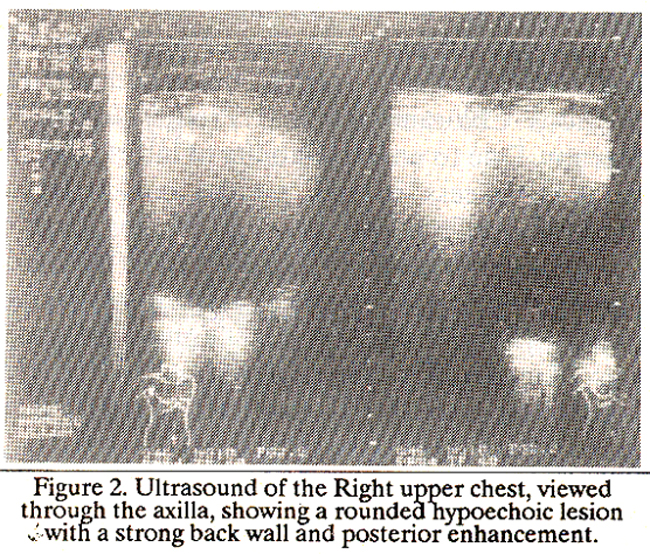
The abdominal structures were normal. Casoni test was positive. The concurrence of imaging and serological results prompted a diagnosis of hydatid cyst of the lung and the patient was put on Albendazole. On follow-up, the patient’s clinical condition had worsened with more pain, weakness and dyspnoea, he also reported an episode of haemetemesis. An X-ray showed an increase in the size of the mass. Bronchoscopy revealed a malignant mass in the right upper lung. Thoracotomy showed this mass to be adherent to the parietal pleura. A right upper lobectomy was done. Histopathology study reported an epidermoid carcinoma with adenomatous metaplasia.
DISCUSSION
Ultrasonography is generally not useful in the diagnosis of lung diseases but, in our experience, peripherally located lesions can, at times, be seen and differentiation of solid from cystic lesions is possible in some cases. We have been successful in visualizing hydatid cysts of the lung, pneumonic consolidation and secondaries. Our failure to detect the consistency in this case might be because of the hypovascularity of the mass2, or the homogeneity of the lesion3. This obviously needs to be examined with more patients and, if a pattern emerges, ultrasound might become an accessory investigation where modalities like CT and bronchoscopy are not available.
REFERENCES
1. O\'Moore, P.V., Mueller, P.R., Simeone, J.F., Saini, S., Butch, RJ., Hahn, P.F., Steimer, E., Stark, D.D. and Ferrucei,J.T. Jr. Sonographic guidance in diagnostic and therapeutic intervention in the pleural space. MR., 1987;149:1.
2. Coleman, B.G., Anger, PH., Mulhern, C.B. Jr., Pollack, N.M., Banner, M.P. and Arenson, R.L. Gray-Scale sonographic spectrum of hypernephromas. Radiology, 1980;137:757.
3. Shirkoda, A., Staab, E.V. and Mittelstaedt, C.A. Renal lymphomas imaged by ultrasound and gallium-67. Radiology, 1980; 137(1 Pt.1):175.
Related Articles
Journal of the Pakistan Medical Association has agreed to receive and publish manuscripts in accordance with the principles of the following committees:


