

Rehana S. Kamal ( Department of Anaesthesiology, The Aga Khan University Hospital, Karachi. )
October 1989, Volume 39, Issue 10
Minor Procedre In Clinical Practice

DIGITAL NERVE BLOCK
1. Anatomy
Each finger and toe is supplied by four nerves, two dorsal and two palmar which run forward along the edge of the digit (Figure 1).
2. Anatomy of Palm.
5ml syringe with a 23-21 Gauge short bevelled needle.
3. Local Anaesthetic solution
2-4ml 1% plain lignocaine. Do not use adrenaline.
4. Indications
Simple operations on the fingers and toes.
5. Contraindications
Extensive injuries or major surgery on the fingers and toes. — Skin infeètion around the base of the finger or teo.
Technique
The digital nerve could be blocked at the base of the finger or toe. Inject 0.5-1ml of solution superficially as well as deeply on either sides of the finger. Avoid injecting more than 1 ml solution at a time, as larger volumes may result in pressure destruction of the blood vessels in the tight area around the base of the finger.
BRACHIAL PLEXUS BLOCK
1. Anatomy
The Brachial Plexus is formed by the fusion of the anterior primary rami of C5, C6, C7, Cg and Ti.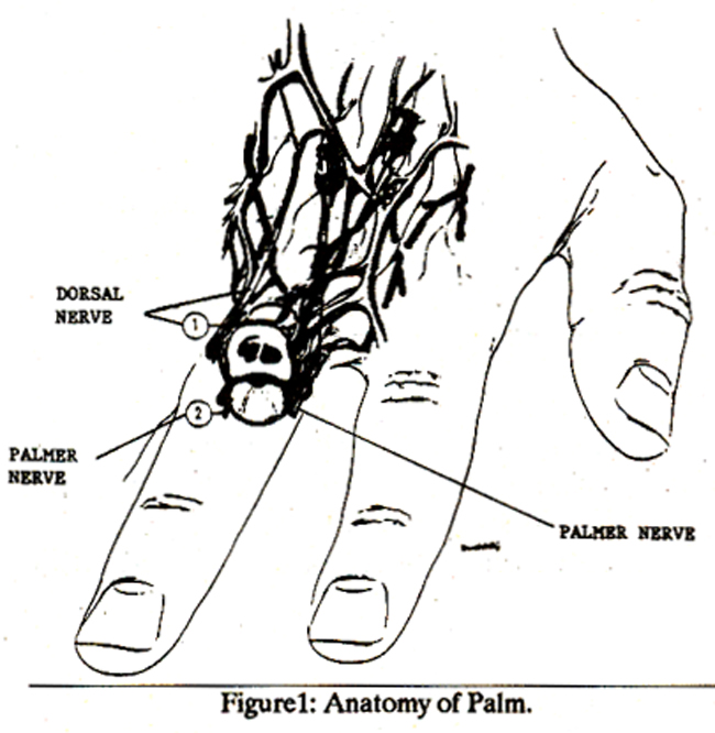
In addition, there is a small contribution from C4 and T2 (Figure 2).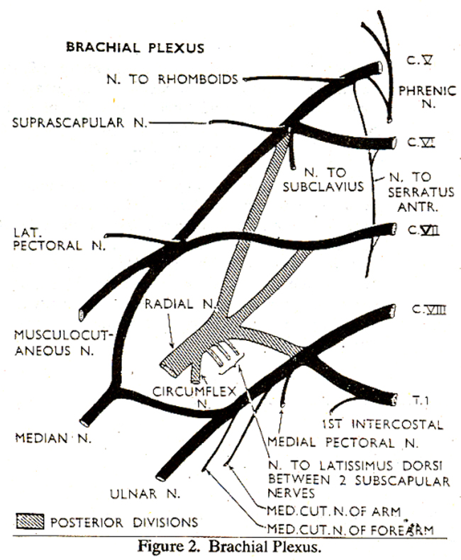
The plexus stretches from the lateral aspect of the cervical vertebral column, downwards and laterally running together with the subclavian artery, between the scalenus anterior and scalenus medius muscles. Across the two muscles is stretched the prevertebral fascia, a tube of prevertebralfascia surrounds the arteries and nerves and continues distally and is called the axillary sheath. This sheath extends to the middle and distal thirds of the arm. The brachial plexus and its terminal branches are contained within this sheath, distributed around the axillary artery (Figure 3).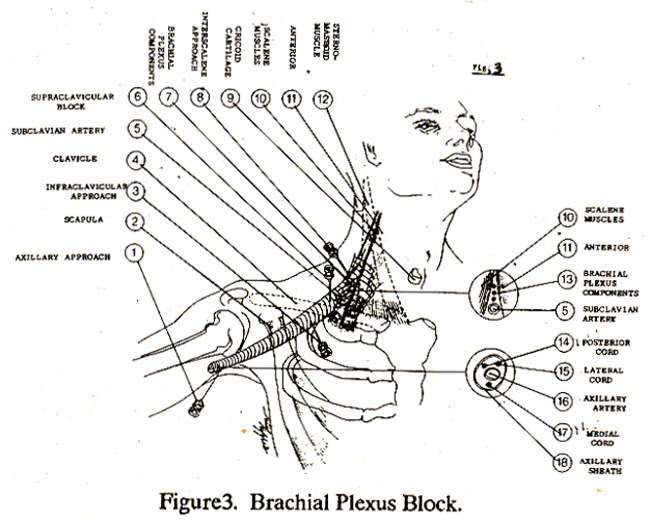
It was always considered a single compartment, but Thompson et al1 demonstrated in cad aver the existence of septa within the sheath, which explains the development of incomplete blocks. The brachial plexus when composed of roots lies between the scalene muscles. The Trunks of the brachial plexus lie in the posterior triangle of the neck. The divisions of the plexus surround the arterybehind the clavicle. It then passes behind the mid-point of the clavicle above the first rib to reach the axillary fossa. Here the brachial plexus joins upto form three main cords which are situated - medially, laterally and posteriorly to the axillary artery. In the axillary fossa the axillary artery, vein and the brachial plexus are enveloped ma common sheath of connective tissue (Figure 4).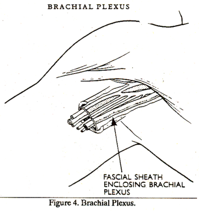
It should be noted that the musculo-cutaneous nerves carrying sensory fibres to the radial side of the forearm leaves the brachial plexus high up in the axilla. It is the most difficult branch to block, especially with the axillary approach.
2. Equipment
20ml syringe with a 21 G needle.
3. Local Anaesthetic
Solutions and dosage. Weight greater than 80kg..
25-30ml of 1.5% ignocaine with adrenaline 1:200,000.
Weight 50-70kg
20-25ml of 1.5% lignocaine with adrenaline 1:200,000.
Weight 40-60kg.
15-20ml of 1.5% lignocaine with adrenaline 1:200,000.
Weight 25-35kg.
14-20ml of 1% lignocaine with adrenaline
1:200,000.
4. Indications
Surgery on the upper limbs. Operations on the hand, forearm and the distal part of the upper arm, especially in out-patient settings. For reduction of fractures, especially if the patient has eaten before the accident.
5. Contraindications
Damage to or disease of the plexus or the distal nerves in the arm.
6. Complications
- Pneumothorax if the supraclavicular approach is used.
- Arterial and Veni puncture.
Technique
Brachial plexus can be blocked either by thr supraclavicular or axillary approach. It is advisable not to try the supraclavicular approach in the out-patient setting because of the complication of pneumothorax. The axillary approach is safer than the supraclavicular approach and is also more acceptable to the patients. With the patient lying supine, the arm is abducted at right angles (i.e.,90°) and the elbow is flexed, the axillary artery is palpated and traced as high in the axilla as possible. Here the plexus lies superflciallyand failure to appreciate this fact, and injecting deeply in the area is the most common cause of failure. The forefinger of the anesthetist’s left hand is placed on the artery, and a short 4-Scm needle is placed slightly above the artery (Figure 5).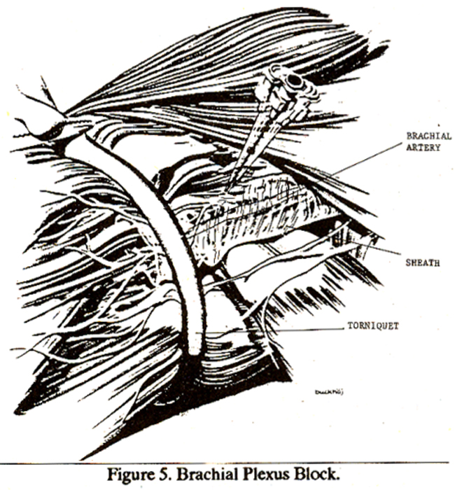
A slight give is felt as the needle enters the sheath, the needle should then pulsate with the arteryindicating that the point of the needle is very near the artery. After careful aspiration the Local Anaesthetic solution is then injected. If parasthesia is elicited, then withdraw the needle slightly before injecting the solution A tourniquet should be applied to the upper arm just below the axilla, to prevent the spread of Local Anaesthetic solution distally. This increases the chances of blocking the musculocutaneous nerve and also may reduce the amount of solution to be injected. The tourniquet should remain for 10 minutes after injection of the solution (Figure 6).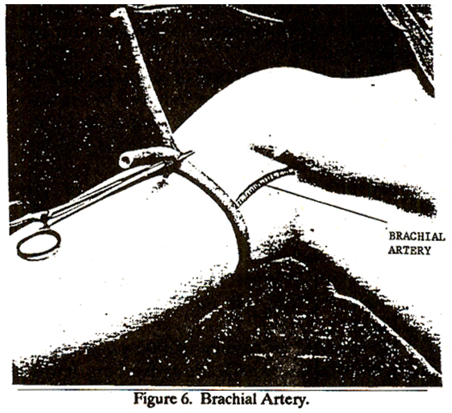
One of the main disadvantages in this technique is the displacement of the needle point, while attaching it to the 20ml syringe. This canbe avoided ERACHIAL by either using a 21g Butterfly or using a flexible extension tubing between the needle and the syringe. Induction time is longer because the Local Anaesthesia has to diffuse to all branches of the plexus, and may take from 20-30 minutes in the adult patients.
REFERENCES
1. Thompson, G.E. and Roire, D.K. Functional anatomyof the branchial plexus sheaths. Anaesthesiology, 1983; 59:117.
2. Vester-Anderson,T., Christiansen, C., Sorensen, M. and Eriksen, C. Perivascular Axillary Block. I. Incomplete Blockade following 40ml 1% mepivacainc with adrenaline. Acta Anaesthesiol. Scand., 1982; 2&519.
Related Articles
Journal of the Pakistan Medical Association has agreed to receive and publish manuscripts in accordance with the principles of the following committees:


