

Mughis Sheerani ( Section of Neurology, Department of Medicine, Aga Khan University Hospital, Karachi. )
January 2005, Volume 55, Issue 1
Review Articles

Introduction and Background
Though medical treatments suffice in almost all types of epilepsy, Epilepsy Surgery is the standard in the management of intractable seizures. There are some misconceptions that lead to underutilization of this treatment, even in the developed countries.1,2 Some of them include:
1. Only temporal lobe seizures are appropriate for surgery.
2. Seizures arising in the dominant hemisphere are not amenable to surgery.
3. There are excessive cognitive or psychiatric risks involved.
4. Focal abnormalities (like spikes) on routine EEG are a prerequisite.
5. Has a low efficacy and high morbidity.
6. Epilepsy surgery evaluation is very costly.3
7. Patient with interictal psychosis cannot undergo surgery.4
Epilepsy is a relatively common disorder in Pakistan. Based on current data, it is estimated that 1.38 million people suffer from epilepsy in the country. In general population, prevalence of epilepsy is estimated to be 9.99 per thousand.5
No comprehensive data is available regarding the numbers of intractable epileptics in our population. Literature suggests that it is common in this part of the world, too. It is reported that almost 240,000 to 320,000 patients of refractory epilepsy in India are potential candidates for epilepsy surgery.6
The extrapolated numbers of potential surgical candidates for Pakistani population, with somewhat same prevalence of epilepsy, would be around 34,000-45,000.
Role of Epilepsy Surgery in developed and underdeveloped countries
Review of literature shows that epilepsy surgery programs are being successfully run in both developed and underdeveloped countries and it is regarded as safe, effective mode of therapy. It is recommended that suitable candidates should be identified early and referred to appropriate centers.2,7,8
A close example is India where several such programs have made a tremendous difference in the life of epileptic patients.7,8 As it is evident, epilepsy surgery requires extensive evaluation and workup. The cost of such treatment remains high, but is comparable to any major surgical procedures, like coronary artery bypass graft. The cost has been estimated in underdeveloped countries too. In India, for example, the out-of-pocket one-time payment for presurgical evaluation for the anterior temporal lobectomy is around 47,000 Indian Rupees (approx. US$1,000). The total direct medical cost of caring for Temporal lobectomy is estimated around 200,000 Indian Rupees (approx. US$4,500). As these patients are mostly young, making them seizure free will help them obtain better employment and improve their quality of life. Therefore, epilepsy surgery is considered a cost effective treatment even for patients in developing countries.3,9
Epilepsy surgery though well-accepted is highly technology dependent. Introducing Epilepsy Surgery in developing countries requires utilization of existing medical infrastructure. This has been achieved in underdeveloped and low socioeconomic countries like India.9,10
Fortunately, Pakistan has several centers that can potentially become Epilepsy surgery centers. They have basic infrastructure and will only need some degree of organization and personnel training to launch an epilepsy program. Following are the broad guidelines to develop such a program that can be modified according to the local requirements and needs.
Epilepsy Surgery Programme
This is a comprehensive, systematic and meticulous diagnostic approach to treat and, if necessary, select the most suitable candidates for Epilepsy surgery. One such program is now being developed at the Aga Khan University Hospital, Karachi.
The team comprises of at least an epileptologist, a neurosurgeon, a neuroradiologist, a neuropathologist and a psychiatrist. Allied heath care professionals include EEG technologists, neuropsychologist, an epilepsy nurse and a social worker.
Equipments and procedures that are utilized includes, Magnetic Resonance Imaging (MRI) Brain, high resolution CT (computerized tomography) scan, angiography and functional imaging (when indicated), EEG (Electroencephalogram), continuous video-EEG (VEEG) monitoring system and computerized spike and event detection most of these tests are available in Pakistan.
MEG (magnetoencehalogram), now an emerging technology, seems to complement EEG for the detection of interictal epileptiform discharges.11 This technology is currently unavailable in Pakistan.
Among non-invasive diagnostic methods, continuous video-EEG monitoring is the most fundamental, cost effective and is also considered a gold standard for the identification of the seizure focus and in turn evaluation for the Epilepsy surgery.12
Video-EEG is now being considered even before placement of a Vagal Nerve Stimulators (VNS) to avoid unnecessary procedures.13
Protocols for Epilepsy Surgery
The protocols are tailored according to the individual patient. Not all studies are prerequisite for every patient. The Epilepsy surgery programme is divided in four phases. The patient has to clear first phase to be considered for the subsequent phase of evaluation and treatment.
Phase I: Initial patient selection.
Phase II: Outpatient pre-surgical evaluation.
Phase III: Inpatient pre-surgical evaluation.
Phase IV: Epilepsy Surgery.
Phase I
Initial Patient Selection
Patient has to undergo detailed outpatient evaluation and pass initial selection criteria before being considered for surgery.
Outpatient Evaluation:
Patient is first evaluated in an outpatient neurology or epilepsy clinic.
Special emphasis is given to a detailed history, especially geared towards: classification of seizure, details about the onset and semiology of the seizures, frequency of the seizures, duration and length of the different antiepileptic drugs (AEDs) used. A detailed psychiatric history and social history, especially details about job responsibilities, are also documented.
Epilepsy clinic also have its own detailed documentation system. This includes the detailed semiology of the seizures, a seizure diary and details of AEDs used and their levels.
Patient selection criteria
These criteria apply to \'non-tumoral\' patients with no circumscribed lesion, such as a low grade glioma or angioma etc. The criteria include:
demonstration of medical intractability (at least one year on three major AEDs with demonstrated therapeutic levels) absence of specified neurological, neurosurgical, severe cardio pulmonary risk and psychiatric contraindications seizures that significantly interfere with patient\'s function surgery offers reasonable opportunity for improvement of function patient and families motivation should be sufficient to perform pre-surgical workup in question patient should, preferably, have at least an IQ greater than 50 for temporal and extratemporal resection and IQ greater than 70 for corpus callosotomy. Chronic psychosis and progressive neurological diseases (except for Rasmussen\'s encephalitis) are excluded generally, age below 50 years (optimal age 5-50 years)
Any above criteria may be altered by consensus in special circumstances.
Phase II
Outpatient pre-surgical Evaluation
Once patient meets above criteria, he or she is selected for an out patient pre-surgical evaluation. These include the following:
1. Routine blood tests including CBC, liver function tests, hepatitis screening and ECG.
2. Routine EEG, if necessary, a sleep deprived EEG.
3. Visual field testing in selected cases.
4. MRI scan (including semi-quantitative measurements of hippocampal volume).
5. SPECT (Single-Photon Emission Computed Tomography) scan and bilateral carotid angiogram in selected cases.
6. Neuropsychological studies including an extensive neuropsychological battery, including, testing for psychometric intelligence, academic achievement, language function, visuo-perceptual, visuo-spatial, and visuo-constructional abilities, short and long term memory for verbal and nonverbal material, auditory perceptual function, sensori-motor abilities, and complex attentional and concentrational skills. Most common one used include: WAIS-R Wechsler memory scale-revised, Warrington recognition memory test and Wisconsin card sorting test. Fluency test and additional examination of verbal and non-verbal memory are undertaken, if necessary.
Non-invasive EEG
All patients undergo 16 channel Video-EEG (VEEG) monitoring in the \'Epilepsy Monitoring Unit\'. This can be done as an outpatient or, if required, as an inpatient procedure. Anti-epileptics are reduced or withdrawn slowly to capture as many seizures as possible. Multiple clinical seizures are recorded and analyzed. An attempt is made to localize the exact focus. At least 4-6 typical seizures are recorded.
The selection for next evaluation proceeds as follows:
1. Invasive recording for Complex partial seizures, without a causal structural lesion.
2. Planning for a focal resection, if all complex partial seizures recorded during noninvasive monitoring, are correlated with the structural lesion.
3. For primary tonic-clonic, tonic, myoclonic, clonic, atonic or combination with each other, are considered for anterior corpus callosotomy.
Phase III
In-patient pre-surgical Evaluation
Once patient fits the initial criteria for possible surgery, he or she is re-admitted to an \'Epilepsy Monitoring Unit\' for further localization of the focus. The testing includes following:
A. Wada Testing
In 1960, Wada and Rasmussen described the intra carotid sodium amobarbital (sodium amytal) test to determine language representation. The goal of this test includes:
1. To establish cerebral representation of the language function.
2. Predicting high risk patients for post-surgical amensitic syndrome
Patient should be at least 4 year old to undergo this test. The interval between the injections is at least 24 hours. Standard 125 mg sodium amytal is used in 5cc solution. Patient must recognize at least 3/5 memory items to fulfill a pass criterion of memory testing.
The results of initial evaluation determine whether patient should undergo further inpatient evaluation.
B. Invasive EEG
Based on the non invasive EEG, patients who will fit the criteria for surgery and/or their pre surgical evaluation reveals equivocal results, is admitted for further invasive VEEG monitoring.
Depending whether focus is extra-temporal or temporal, following decisions, regarding invasive monitoring, are taken:
1. Invasive EEG protocols when extra-temporal lobe epilepsy is suspected
2 Invasive EEG protocols when temporal lobe temporal epilepsy is suspected
1. Invasive EEG Protocols when Extratemporal Epilepsy is suspected:
When extra-temporal focus is identified and needs further localization, following approach is usually undertaken:
A. When non-invasive evaluation is not lateralized:
Bilateral depth electrodes in temporal and frontal lobes are used for further lateralization.
Additional depth/ subdural electrode can be utilized, if needed, for localization.
B. When non-invasive evaluation is lateralized/ localized:
Subdural mats/ strips, as indicated, for further localization and functional evaluation are used.
C. For suspected frontal lobe epilepsy:
Bilateral orbital frontal, supplementary motor depth electrodes, bilateral frontal convexity and inter-hemispheric subdural grids are usually used for further localization.
2. Invasive EEG Protocols when Temproal Lobe Epilepsy is suspected:
When non-invasive EEG point towards temporal lobe epilepsy (TLE) that needs further localization, one of the following approach is undertaken:
A. Placement of bilateral hippocampal occipital depth electrodes and bilateral temporal subdural strip electrodes.
B. Placement of orbital, hippocampal, amgydalar electrodes via coronal approach.
C. When temporal versus frontal seizure onset is in question, bilateral temporal and frontal depth electrodes and subdural strip recording is utilized.
Varieties of Electrodes
Following different types of electrodes that commonly utilized depending on the case:
Strip electrodes, Forman ovale electrodes, depth electrodes/SEEG(stereotacticdepth electroencephalography) or electrode grids. Among these subdural grids seems to be the most informative.
Sub-dural Grids
Sub-dural grids with continuous video-EEG monitoring are performed in patients
1. When there is inconsistent spike localization or morphology.
2. When there is inconsistency between spike and seizure.
3. When there are bilateral spikes without clear predominant lateralization.
4. When there are bilateral seizure discharges without clear lateralizing predominance.
Subdural grids are avoided in patients with localized swelling or edema.
Risk and Complications of Subdural Grids
Patients with intractable epilepsy can benefit from subdural invasive monitoring procedures but does entail definite, but acceptable, risks. Risks and complications are usually transient and include: cerebrospinal fluid leaks, cerebral edema, subdural hematoma, postprocedural intracerebral hematoma, hypertrophic scars, infections, including osteomyelitis and superficial wound infections. It has been reported that blood loss during the procedure and the amounts of subsequent transfusions correlates directly with the size and number of electrodes on the grids.14
Increased complication rates have been reported in patients with left-sided grid insertion and longer monitoring with a greater number of electrodes (especially more than 60 electrodes).14,15
Phase IV
Once the focus is localized and mapped, best surgical procedure is planned by the team. As temporal lobe epilepsies are the most common to undergo surgical treatment, following are some points to be noted:
Important Facts about temporal lobe Epilepsies
Well-selected TLE patients can derive maximal benefit from anterior temporal lobectomy (ATL) after a noninvasive presurgical evaluation. This finding is of great significance for the creation of epilepsy surgery programs in developing countries which can be very cost-effective for the patients in the long run.3
Bitemporal foci can also be evaluated for epilepsy surgery. In these patients, invasive monitoring to pursue possible surgical therapy may be justified only when some lateralizing features are found in other noninvasive assessments.12
Surgical Techniques
Several types of procedures and surgeries are available once the type and focus of the seizures is Table 1.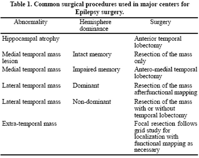
Common surgical procedures used in major centers for Epilepsy surgery.
Decision on non-invasive testing
When noninvasive testing, e.g. MRI or Video-EEG, provides positive results, surgery is considered according to the site of the abnormality. Table 1 shows the common surgical procedures used in major centers.20
Decision on invasive testing
Table 2 illustrates the way surgical decision is undertaken depending on the epileptogenic focus as determined on the invasive testing.
Hemispherictomy
Hemispherictomy is considered in seizures originating from an injured hemisphere causing multifocal seizures. Invasive testing is usually not required. It is considered only for non-dominant hemisphere where speech and memory function should not reside. Patient with hemiplegia and no useful hand function of that limb can also be considered for this kind of surgery.
Corpus Callosum Section
Corpus callosum section is considered for intractable atonic or major motor seizures causing marked impairment of lifestyle. These patients are not candidates for focal resection. Invasive monitoring is not usually performed.
Table 2. Surgical procedures for Epilepsy surgery depending on EEG.
The EEG, ideally, should show a generalized onset, preferably, anterior frontal locations. Speech and handness should reside in the same hemisphere. Standard procedure is to section anterior 2/3 of the corpus callosum through the right frontal approach. If this proves inadequate, the patient may require complete callosotomy.
Multiple Subpial Transections
MST is a novel surgical approach for certain refractory epileptics. The aim of the procedure is to cut horizontal intracortical fibers. The vertical afferent and efferent connections and vasculature is spared. It is hypothesized that horizontal fibers are responsible for the synchrony noted in the seizure activity. The transections disrupt the synchrony there by controlling the spread of the seizure activity. These transections have been applied in relatively sensitive areas like, pre and post central cortex, without producing any functional deficits.
Vagal nerve stimulation
This procedure is used for patients who have been excluded for any resective procedure. There is up to 50% reduction in the seizure control.3 Patient and the care givers can actually stop a seizure, if they catch it early. The procedure is relatively safe with minimal complications.
Other surgical techniques
Extratemporal epilepsies (EE) can undergo lesionectomies, disconnecting procedures, polectomies and/or lobectomies, corticectomies and anatomical hemispherectomy. In several studies, surgical outcomes range from very good to at least worthwhile.20
Decision on invasive testing
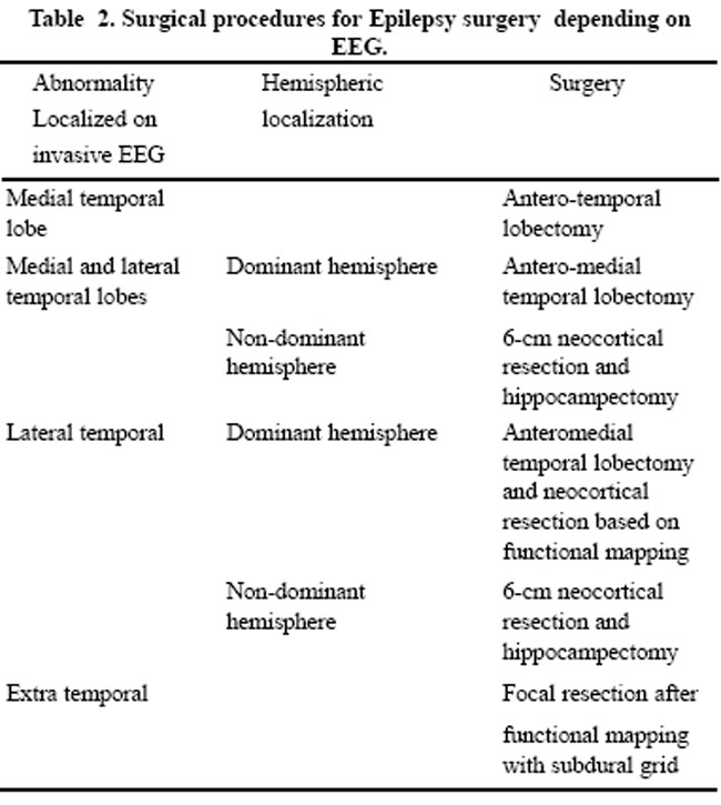
Table 2 illustrates the way surgical decision is undertaken depending on the epileptogenic focus as determined on the invasive testing.
Hemispherictomy
Hemispherictomy is considered in seizures originating from an injured hemisphere causing multifocal seizures. Invasive testing is usually not required. It is considered only for non-dominant hemisphere where speech and memory function should not reside. Patient with hemiplegia and no useful hand function of that limb can also be considered for this kind of surgery.
Corpus Callosum Section
Corpus callosum section is considered for intractable atonic or major motor seizures causing marked impairment of lifestyle. These patients are not candidates for focal resection. Invasive monitoring is not usually performed.
The EEG, ideally, should show a generalized onset, preferably, anterior frontal locations. Speech and handness should reside in the same hemisphere. Standard procedure is to section anterior 2/3 of the corpus callosum through the right frontal approach. If this proves inadequate, the patient may require complete callosotomy.
Multiple Subpial Transections
MST is a novel surgical approach for certain refractory epileptics. The aim of the procedure is to cut horizontal intracortical fibers. The vertical afferent and efferent connections and vasculature is spared. It is hypothesized that horizontal fibers are responsible for the synchrony noted in the seizure activity. The transections disrupt the synchrony there by controlling the spread of the seizure activity. These transections have been applied in relatively sensitive areas like, pre and post central cortex, without producing any functional deficits.
Vagal nerve stimulation
This procedure is used for patients who have been excluded for any resective procedure. There is up to 50% reduction in the seizure control.3 Patient and the care givers can actually stop a seizure, if they catch it early. The procedure is relatively safe with minimal complications.
Conclusion
Epilepsy surgery is a proven and effective treatment for intractable seizures. In long term, it is also cost effective, even in underdeveloped countries like Pakistan. Evaluation through a \'Comprehensive Epilepsy Surgery Programme\', before surgery, improves the likelihood of success and reduces the risk of complications.
References
1. Benbadis SR, Heriaud L, Tatum WO, Vale FL. Epilepsy surgery, delays and referral patterns-are all your epilepsy patients controlled? Seizure 2003;12:167-70.
2. Spencer SS, David King D. Surgical options in the management of the epilepsies in adults and older children. Neurologist 1997;3:2-12.
3. Campos MG, Godoy J, Mesa MT, Torrealba G, Gejman R, Huete I. Temporal lobe epilepsy surgery with limited resources: results and economic considerations. Epilepsia 2000;41 (Suppl 4 ):S18-21.
4. Marchetti RL, Fiore LA, Valente KD, Gronich G, Nogueira AB, Tzu WH. Surgical treatment of temporal lobe epilepsy with interictal psychosis: results of six cases. Epilepsy Behav 2003;4:146-52.
5. Khatri IA, Iannaccone ST,Ilyas MS, et. al. Epidemiology of epilepsy in Pakistan: review of literature. J Pak Med Assoc 2003;53:594-7.
6. Kar AM , Garg RK , Verma R. Refractory epilepsy: diagnosis and management. J Indian Med Assoc 2002;100:290-4.
7. Shukla G , Bhatia M , Singh VP , Jaiswal A , Tripathi M , Gaikwad S, et al. S Successful selection of patients with intractable extratemporal epilepsy using non-invasive investigations. Seizure 2003;12:573-6.
8. Bhatia M , Singh VP , Jain S , Gaekwad S , Bal CS , Sarkar C , et al. Epilepsy surgery in India: All India Institute of Medical Sciences experience. J Assoc Physicians India 1999;47:492-5.
9. Rao MB, Radhakrishnan K Is epilepsy surgery possible in countries with limited resources? Epilepsia 2000;41(Suppl 4):S31-4.
10. Williamson PD, Jobst BC. Epilepsy surgery in developing countries. Epilepsia 2000;41 (Suppl 4):S45-50.
11. Barkley GL, Baumgartner C. MEG and EEG in epilepsy. J Clin Neurophysiol 2003;20:163-78.
12. Cascino GD. Video-EEG monitoring in adults. Epilepsia 2002;43 (Suppl 3):80-93.
13. Attarian H, Dowling J, Carter J, Gilliam F. Video EEG monitoring prior to Vagal nerve stimulator implantation. Neurology 2003;61:402-3.
14. Onal C, Otsubo H, Araki T, Chitoku S, Ochi A, Weiss S, et al. Complications of invasive subdural grid monitoring in children with epilepsy. J Neurosurg 2003;98:1017-26.
15. Hamer HM, Morris HH, Mascha EJ, Karafa MT, Bingaman WE, Bej MD, et al. Complications of invasive video-EEG monitoring with subdural grid electrodes. Neurology 2002;58:97-103.
16. Sinclair DB, Aronyk K, Snyder T, McKean J, Wheatley M, Bhargava R, et al. Pediatric temporal lobectomy for epilepsy. Pediatr Neurosurg 2003;38:195-205.
17. Holmes MD, Miles AN, Dodrill CB, Ojemann GA, Wilensky AJ. Identifying potential surgical candidates in patients with evidence of bitemporal epilepsy. Epilepsia 2003;44:1075-9.
18. Spencer SS, When should temporal-lobe epilepsy be treated surgically? Lancet Neurol 2003;1:375-82.
19. Pomata HB, Gonza¡lez R, Bartuluchi M, Petre CA, Ciraolo C, Caraballo R, et al. Extratemporal epilepsy in children: candidate selection and surgical treatment. Childs Nerv Syst 2001;16:842-50.
20. Holmes MD, Dodrill CB, Ojemann GA, Wilensky AJ, Ojemann LM. Outcome following surgery in patients with bitemporal interictal epileptiform patterns. Neurology 1997;48:1037-40.
Journal of the Pakistan Medical Association has agreed to receive and publish manuscripts in accordance with the principles of the following committees:


