

Salimuddin Aziz ( PMRC Research Centre, Jinnah Postgraduate Medical Centre, Karachi. )
Syed Ejaz Alam ( PMRC Research Centre, Jinnah Postgraduate Medical Centre, Karachi. )
January 1990, Volume 40, Issue 1
Original Article

Abstract
In a retrospective study, case records of 1061 patients treated with antituberculosis drugs were examined to assess the criteria for diagnosis in each case. Seventy six percent had sputum examined for A.F.B. and 45% had mantoux test done. Five hundred and eighty one (55%) were diagnosed on radiology alone, while 262 (25%) had positive sputum for A.F.B. Only 50(5%) of cases had positive Mantoux in addition to positive sputum and radiological changes. Practical significance of this practice is discussed (JPMA 40:14, 1990).
INTRODUCTION
In a previous study1 it was observed that a group of patients were being treated with antituberculosis drugs, when they were sputum (Direct smear and culture) and Mantoux negative. Diagnosis in these cases was considered suspect. Since pulmonary tuberculosis is diagnosed rather frequently in this part of the world, and treatment is long drawn and expensive, it was felt that criteria of diagnosis should be evaluated in the light of Mantoux test, Sputum studies, X-Ray and histology to determine the accuracy of diagnosis and to document the drugs used, their compliance and outcome of the treatment.
MATERIAL AND METHODS
Three years (1985-1987) case records of pulmonary tuberculosis patients who had received anti-tuberculosis therapy in three different institutions of Karachi, i.e., a teaching hospital, a chest clinic and a private hospital were retrospectively analysed to determine the criteria used for the diagnosis, combinations of drugs used, their duration, compliance and eventual outcome. To extract maximum information, the data of.all age groups and both sexes was recorded on a standardised proforma. A total of 1061 patients were included in the study, of these 319 belonged to hospital, 413 to the chest clinic and 329 to a private hospital.
RESULTS
One thousand sixty one (731 male, 330 female) patients were studied, their mean age was 36 years with a range of 2-80 years.
Teaching Hospital
A total of 319 (197 male, 122 female) cases were analysed whose mean age was 37 years with a range of 10-80 years. Sputum was examined in 233(73%) cases of which 91(39%) were positive for A.F.B. Mantoux was performed in 63(20%) of which 27 (43%)were secorded as positive. Two hundred and six (65%) were diagnosed on X-Ray alone, while 19 (6%) had positive Mantoux with X-Ray change. Sputum was positive alongwith radiological change in 91 (29%) while 8 (3%) had Mantoux positive in addition (Table I).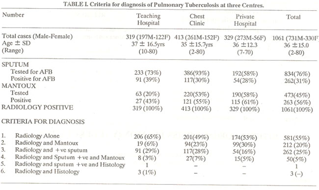
Chest Clinic
Total of 413 (261 male, 152 female) cases were studied with an average age of 35 years and a range of 2-80 years. Sputum was examined in 386 (93%) cases and 117 (30%) were found pasitive.
Mantoux was done in 220 (53%) and documented as positive in 121 (55%). Two hundred one (49%) were diagnosed on X-Ray alone while 94 (23%) had positive Mantoux with X-Ray changes. Sputum was positive, along with X-Ray changes in 117 (28%) while Mantoux was positive in 27 (7%) (Table I).
Private Hospital
A total of 329 (273 male, 56 female) with mean age of 36 years with a range of 7-70 years were analysed. Sputum was examined in 192 (58%) and 54 (28%) were found positive. Mantoux was done in 190(58%) and found positive in 115(61%). One hundred seventy four (53%) were diagnosed radiologically alone, while 99 (30%) had positive Mantoux with radiological changes. Sputum was positive in 54(16%), while Mantoux was positive in 15(6%) in addition.
Overall 581 (55%) of 1061 were diagnosed on radiology alone while 212 (20%) had positive Mantoux with radiological change. Sputum was positive in 262 (25%) while Mantoux was positive in 50(5%) in addition.
Follow up data was available at the chest clinic and private hospital. Of these 283 (38%) completed the treatment (clinic 35%, P. hospital 42%) and 59(8%) were continuing treatment. The defaulters were 322(43%) and amongst 76 (10%) it was unknown (Table II).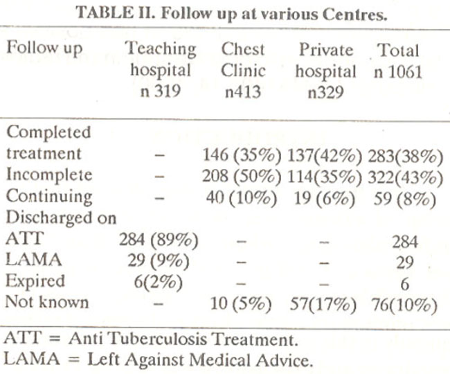
DISCUSSION
Many patients seek help for respiratory ailments and since tuberculosis is rather common in our country, a fair number are put on antituberculosis drugs. In the present study more than half of those actually being treated for pulmonary tuberculosis were diagnosed on radiology alone. It seems that it is still widely believed that pulmonary tuberculosis can be diagnosed by X-rays alone but a number of studies confirm that no radiological picture is typical of tuberculosis, and lesions of pulmonary tuberculosis can take almost any form2. In addition inter individual3-5 and intra individual6,7 errors are well recognised, confirming that purely radiological criteria cannot give satisfactory evidence of tuberculosis in an individual patient8. Sputum positivity is an essential component for defining a case” of tuberculosis according to the World Health Organisation. In the present study only 76% of patients were subjected to sputum examination (Table I) and only 25% were A.F.B. positive leaving large numbers (75%) of “suspects" with either abnormal X-Ray or positive mantoux in addition. Sputum examination if property done, will yield 76.4 to 83 percent positive results9,10 two overnight specimens have a better yield than 2 spot specimens. It is true that in patients with early or suspected tuberculosis infection or with disseminated fibrotic lesions tuberculous bacillus may be so scanty that direct smear microscopic examination is not sufficiently sensitive to detect organisms, but when the bacillus numbers more than 100,000/ ml then it is consistently detected11,12. It was discovered that in the absence of sputum no other efforts, like laryngeal swabs or gastric washing were done to clinch diagnosis. Mantoux was the least performed of all investigations, especially in the teaching hospital (20%). Many physicians do not consider it a worthwhile test, since vast majority of adult population is Mantoux positive13 but in spite of this limitation it is a very helpful test, specially in excluding tuberculosis except under special circumstances and should form an essential routine test in the investigations.
This study confirms the original observation that due care is not taken to confirm the diagnosis before embarking on prolonged expensive treatment. Too much reliance is put on radiology, which though very sensitive is not specific for tuberculosis. This practice is not only the cause Of waste of scarce resources but also the correct diagnosis is missed in many patients. Special care should, therefore, be taken in collection of sputum, smear preparation, selection of the right part of the sputum and careful scanning of slides for better yield. Every effort should be made to employ all means to confirm the diagnosis bacteriologically. Trained people should be attached to a tuberculosis unit (such staff is easy to train) and even non- specialised staff are capable of carrying out satisfactory smear microscopy. The limited resources make it mandatory that these should be used judiciously. Follow up in the teaching hospital
leaves a lot to be desired (Table II) and better method needs to be devised. Chest clinic is well organised, follow tip is comprehensive though default is high. This institution comes out best in comparison to others.
ACKNOWLEDGEMENT
Our thanks are due to the heads of Departments of all the three institutions for allowing us access to their records, and also to their staff for cooperation.
REFERENCES
1. Aziz, S. and Haq, 0. The Mantoux in pulmonary tuberculosis.Tubercle, 1985; 66:133.
2. American Lung Association. Diagnostic standard, classification of tuberculosis and other mycobacterial diseases. NewYork, Am. Lung Assoc., 1974, p.22.
3. Garland, L.H. Studics on the accuracy of diagnostic procedures.AJR., 1959; 82:25.
4. Garland, L.H. On the scientific evaluation of diagnostic procedures. Radiology, 1949; 52:309.
5. Newell, R.R., Chamberlain, W.E. and Rigler, L. Descriptive classification of pulmonary shadows. Revelation of unreliability in roentgenographic diagnosis of tuberculosis. Am. Rev. Tuberc., 1954; 69:566.
6. Cochrane, A.L. and Garland, LII. Observer error in interpretation of chest films; an International investigations. Lancet, 1952; 2:505.
7. Nakaamura, K et al. kekau. 1970; 45 : 121. Quotcd by Toman, K. in Tuberculosis, case finding and chemotheracy. Geneva, World Health Org., 1979, p.30.
8. World Health Organization, WHO Expert committee on tuberculosis. WHO Tech. Rep. Ser., 1974; 552:8.
9. Nagpaul, D.R, Savie, D.M., Rao, K.P. et al. Case-finding by microscopy. Bull. Int. Union Tuberc., 1968; 41: 148.
10. Andrew, R.H. and Radhakrishna, S. A comparison of two methods of sputum collection in the diagnosis of pulmonarytuberculosis.Tubercle, 1959; 40: 155.
11. Carvalho, A. Zeitschrift fiir tuberculous Und erkrankungen der Thoraxorgane (1932). 63: 305. Quoted by Chandrasekher, S. Text book of tuberculosis. Vikas Publishing House 1981 p. 2020.
12. Crukshank, D.B., Sellors, T.H. and Livingstone, J.L. ed. Modern practice in tuberculosis. London, Butterworth, 1952; p.62.
13. Kalota, J. Epideniiological situation of tuberculosis in Pakistan national prevalence Survey 1974-78.
Journal of the Pakistan Medical Association has agreed to receive and publish manuscripts in accordance with the principles of the following committees:


