

Zulfiqar A. Bhutta ( Department of Paediatrics, The Aga Khan University Hospital, Karachi. )
Shehla H Naqvi ( Department of Paediatrics, The Aga Khan University Hospital, Karachi. )
June 1991, Volume 41, Issue 6
Case Reports

Flavobacterium meningosepticum is a gram negative, opportunistic organism which has been implicated in a number of outbreaks of sepsis and meningitis in newborn nurseries1-4. The infection is usually associated with a very high mortality3,4. F. meningosepticum is usually resistant to most of the commonly used antibiotics5 and successful therapy has posed problems. We present a case report of F. meningosepticum meningitis caused by a multiply drug resistant strain of the organism.
CASE REPORT
A four day old newborn male was transferred to Aga Khan University Hospital (AKUH) from another hospital with a history of left side seizures for 48 hours. The baby was born at full term, after spontaneous vaginal delivery to a primipara who was hypertensive during pregnancy. Birth weight was 2.3kg. There were no immediate problems, but two hours after birth the baby became jittery. Blood glucose was 32 mg/dl and serum calcium was 7.5 mg/dl. Glucose and calcium gluconate were given intravenously. Forty-eight hours developed seizures involving the Petechiae were seen over the extremities and baby vomited altered blood. Platelet count was 25 x 109/L but prothrombin time and partial thromboplastin time was not prolonged. Diazepam was used to control seizures and as sepsis was suspected, cefotaxime and amikacin were given intravenously. The patient had an apneic episode following which he was transferred to AKUH. At AKUH the baby was found to be hypotonic with no spontaneous movements. B.P. was normal 73/50 mmllg, HR 170/mm, RR 32/mm. Temp. was 37°C. Skin perfusion was poor. A loud systolic (2/6) murmur was present. Apneic episodes of 20-25 seconds occurred every 5-10 minutes. Liver was palpable two cm below the right costal margin. Length and head circumference were at 25th percentile while weight was at the 5th percentile. The baby was assessed to be small for gestational age with postnatal sepsis and patent ductus arteriosus. Lumbar puncture was done after infusion of platelets and fresh frozen plasma. CSF cell count and chemistry could not be done due to the small sample obtained. Cefotaxime and Amikacin were continued after blood and CSF cultures. Serum bilirubin had risen to total of 24.3 mg/dl with 20.3 mg/dl unconjugated fraction. Exchange transfusion was performed and the baby was mechanically ventilated. Abdominal distension was a persistent problem but no radiologic signs of necrotizing enterocolitis were seen. Blood and CSF cultures remained negative, but as they were obtained after the institution of antimicrobial therapy, antibiotics were continued. Cranial ultrasonography revealed a left subependymal hemorrhage on day of admission. Seizure continued despite adequate anticonvulsant therapy. Platelets remained low. Cranial computerized tomography was performed on seventh hospital day and showed dilatation of ventricular system with contrast enhancement suggestive of ventriculitis. Ventricular CSF was examined (Table)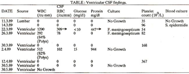
and found to be abnormal with Grain negative bacilli seen on gram stain. Initially the organism which was cultured from CSF was suspected to be Pseudomonas and therapy was changed to carbenicillin and tobramycin. These were discontinued when the isolate was identified as F. meningosepticum which was susceptible to ceftizoxime and ofloxacin and resistant to carbenicillin, gentamicin, tobramycin, amikacin, ceftriaxone, cefotaxime, polymyxin B and rifampicin on disc diffusion/testing. As ceftazidime was not readily available, rifampicin was started in a dosage of 15 mg/Kg/day I.V Q 12H alongwith ceftazidime 100 mg I.V + Q 12H. Ventricular CSF was reexamined 48 hours after starting rifampicin and ceftazidime therapy, and againyielded F. meningosepticum. Ceftizoxime was substituted for ceftazidime and rifampicin at this stage (five days after diagnosis of ventriculitis). Severe recurrent seizures which had persisted, ceased four days after starting ceftizoxime and platelet count which was persistently low, became normal (168000/cumm). Ventricular CSF was found to be sterile one week after the initiation of this therapy. The patient started feeding and gaining weight. Treatment was continued for two more weeks after documenting sterilization of CSF when rifampicin and ceftizoxime were discontinued. The progressive improvement in laboratory studies is shown in table. Patient was discharged home however, with instructions to return for repeat CSF examination. The parents did not bring him back for one and a half months at which time he was found to have obvious hydrocephalus. Head circumference had increased to 44 cm from 33.5 cm at discharge. His neurologic examination showed increased tone and marked hyperreflexia. Sunset appearance of eyes was present. He had also gained weight from 2.15 Kg at discharge to 4.5 kg. A ventriculoperitoneal shunt was placed and child was discharged after 9 days hospitalization. Ventricular CSF cultured at this time was sterile. He has subsequently been reviewed at 9 months of age and has significantly developmental delay with increased tone in both lower limbs.
DISCUSSION
Flavobacterium meningosepticum is usually a water borne organism and has been known to proliferate in wash basins and aqueous antiseptic solutions6, leading to hospital outbreaks. There have been very few cases reported from the South Asian subcontinene7. The Aga Khan University Hospital in Karachi has neonatal tertiary care facilities and consequently receives patients from a large referral population. Over the last three years 88 cases of culture proven cases of neonatal septicemia and/or meningitis were admitted to the newborn care services at AKUH. Flavobacterium meningosepticum was isolated in two cases (2.3%). Both the cases were referred from outside facilities and were not associated with outbreaks in the referring hospitals. Successful treatment of the F. meningosepticum infection poses a number of difficulties as the antibiotic susceptibility pattern is unique. The organism is usually resistant to conventional first line antibiotics used in the neonatal period such as penicillin, gentamicin, ampicillin and kanamycin. However, therapeutic success has been described with intraventricular erythromycin8 intraventricular with intravenous rifampicin9 and recently with intravenous trimethoprim-sulfa5. Our patient was referred from outside and did not have any identifiable risk factors for sepsis other than intrauterine growth retardation. The baby developed signs of meningitis within 48 hours after birth suggesting early acquisition of the infection. Although the organism was resistant to rifampicin and ceftazidime on disc diffusion testing, ceftizoxime was not readily available and an interim therapy was initiated with these drugs. Ceftizoxime is a newer third generation cephalosporin and has been used in the therapy of gram positive and negative meningitis10. However, to our knowledge, there are no case reports of successful treatment with ceftizoxime in multiply drug resistant meningitis. We were able to sterilize the CSF in our case, but the baby developed obstructive hydrocephalus and neurological sequelae. This is understandable in view of the delay in initiation of appropriate therapy (15 days from onset of illness). Infection with F. meningosepticum had also been associated with a high incidence of neurological sequelae3,4. Third generation cephalosporins have revolutionized the therapy and outcome of gram negative meningitis and our experience indicates that ceftazidime may also prove useful in the treatment of F. meningosepticum sepsis and meningitis also which have posed severe problems in therapy todate.
REFERENCES
1. Abrahamsen, T., Finne, P. H. and Lingaas, E. Flavobacterium meningosepticum infections in a neonatal intensive care unit. Acta Paediatr. Scand., 1989; 78: 51.
2. Cabrera, H. A. and Davis, 0. H. Epidemic meningitis of the newborn caused by flavobacteria. I. Epidemiology and bacteriology. Am. J. Dis. Child., 1961; 101: 289.
3. Ratner, H. Flavobacterium meningosepticum. Infect. Control, 1984; 5: 237.
4. George, it M., Cochran, C. P. and Wheeler, W. E. Epidemic meningitis of the newS horn caused by flavobacteria. II. Clinical manifeatationa and treatment. Am. 3. Dia. Child., 1961; 101: 2%.
5. Thong. 84. L., Puthucheary, S. D. and Lee, EL Flavobacterium meningosepticum infection; an epidemiological study in a newborn nursery. .1. Clin. Pathol., 1981; 34: 429.
7. Golcul, B. N., Chandramukhi, A., Ravikumar, R. and Aroor, S. Flavobacterium meningosepticum meningitia in a neonate. IndianJ. Pediatr., 1989; 56: 524.
8. Maderazo, E. 0., Bassaria, H. P. and Quintiliani, R. Flavobacterium meningosepticum meningitia ins newborn infant. 3. Pediatr., 1974; 85: 675.
9. Lee, EL, Robinson, M.J., Thong, M.L and Puthuchesry, S.D. Rifamycin in neonatal liavobacteria meningitis. Arch. Dis. Child., 1976; 51: 209.
10. Cable, D., Edralin, 0. and Overturf, 0. D. Human cerebrospioal fluid phar macokinetica and treatment of bacterial meningitis with ceftizoxime. 3. Antimicrob. Chemother., 1982: 10 (Supple c.): 121.
Related Articles
Journal of the Pakistan Medical Association has agreed to receive and publish manuscripts in accordance with the principles of the following committees:


