

Riffat Bano ( Department of Obstetric and Gynae, Aga Khan Hospital for Woman, Karimabad, Karachi. )
Ammara Mushtaq ( Medical Student, Dow Medical College, Dow University of Health Sciences, Karachi. )
Mehreen Adhi ( England Eye Center, Tufts Medical Center and Tufts University School of Medicine, Boston, USA. )
Nousheen Asim ( Department of Obstetric and Gynae, Aga Khan Hospital for Woman, Karimabad, Karachi. )
Noureen Afzal ( Department of Paediatrics, Aga Khan Hospital for Woman, Karimabad, Karachi. )
November 2013, Volume 63, Issue 11
Short Reports

Methods and Results
The hospital-based descriptive study was carried out over a period of two years, at the Aga Khan Hospital for Women, Karachi, from January 2009 to December 2010. All pregnant women who delivered at the hospital between 32-41weeks of gestation were included. Very premature babies (gestational age < 32weeks) and those with congenital anomalies were excluded from the study. All SGA cases were diagnosed antenatally by clinical examination and ultrasound. For SGA foetuses, maternal age, gestational age, mode of delivery and birth-weight were recorded. The modes of delivery were divided into three groups: spontaneous vaginal deliveries, instrumental deliveries and Caesarean sections. Birth-weight of each baby was measured without clothes to the nearest 100 gram on a baby-weighing scale within one hour of birth. Baby scale was calibrated before weighing each baby. Based on the birth-weight, the SGA cases were divided into three groups: 1-1.5kg, 1.6-2 kg, 2.2-2.5 kg. Gestational age was estimated by ultrasonography and was categorised as delivered at less than 34 weeks, 34-37 weeks and greater than 37 weeks. Maternal age was recorded at the time of delivery and categorised as less than 20 years, 20-40 years and greater than 40 years. Of the 6024 deliveries conducted during the study duration, 722(11.98%) caried SGA foetuses. The foetal weight was 2.1-2.5 kg for 616(85.13%) cases (Table).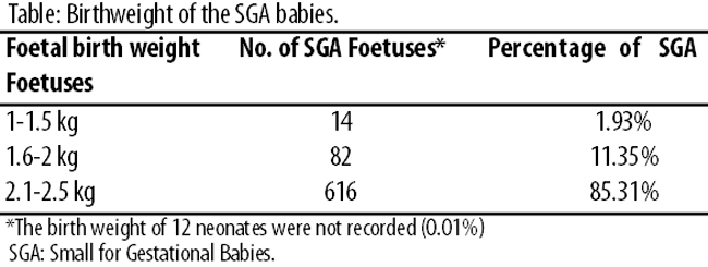
The maternal age in 588(81.44%) cases was between 20-40 years. It was less than 20 years in 121 (16.75%) cases, and over 40 years for 13(1.80%). The gestational age in 406 (56.23%) cases, was 34-37 weeks. It was less than 34 weeks in 21(2.90%) cases, and more than 37 weeks in 295(40.85%).
Correlating SGA with mode of delivery, 170(53.96%) cases were delivered by vaginal route (Figure-1).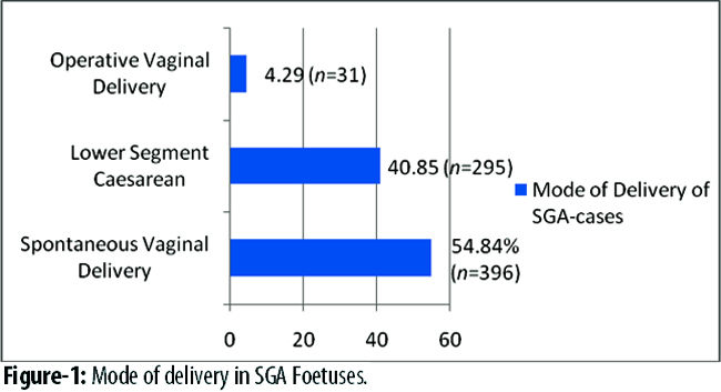
The frequency of SGA deliveries in 2010 was 22% more than in 2009 (13.13% vs 10.76%) (Figure-2).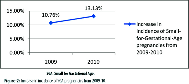
Abstract
The objective of the study was to determine the frequency and outcome of Small for Gestational Age (SGA) foetuses, and its association with the mode of delivery, foetal birthweight, maternal age and gestational age. It was a hospital-based descriptive study, conducted at Aga Khan Hospital for Women, Karachi, from January 2009 to December 2010. Mode of deliveries, foetal birthweight, maternal age and gestational age of SGA foetuses were recorded and analszed. Of a total of 6024 deliveries during the study period, 722(11.98%) had SGA foetuses. Maternal age in such cased ranged between 20-40 years in 588(81.44%), while 406(56.23%) of these deliveries were in between 34-37 weeks of gestation. Besides, 396(54.84%) cases were delivered vaginally. The incidence of SGA increased from 2009 to 2010 (13.13% vs 10.76%). The frequency of SGA foetuses tends to be significantly higher in Pakistan compared to the Western world. Prenatal care and better management can potentially avoid the occurrence of SGA deliveries, perinatal morbidity and mortality.
Keywords: Small-for-gestational-age, Mode of delivery, Foetal outcome, Birthweight, Gestational age.
Introduction
Small for Gestational Age (SGA) foetuses are defined as foetuses having weight less than 10th percentile for its gestational age and abdominal circumference less than 2.5th percentile. Alternatively, SGA foetuses have birthweight and/or length two standard deviations below the mean for gestational age.1
SGA infants are at a greater risk of mortality than Appropriate-for-Gestational (AG) infants.2 They are also at increased risk of hypospadias, bronchopulmonary dysplasia, lower placental and foetal weight, obesity, hypoglycaemia, polycystic ovary syndrome, neurodevelopmental disability, stillbirths, among many others.3-9
In Pakistan, a review in 2004 reported 3-10% pregnancies are SGA foetuses.10 Intrauterine growth restriction (IUGR)- a class of SGA has a frequency of 23.8% all over the world, with 75% of them in Asia.11 In Pakistan, the incidence of IUGR alone is 25%.11 Data from a tertiary-care hospital in Karachi in 2011 identified foetal growth restriction as among the most common causes of stillbirth.12
Although a number of studies previously have reported frequency of SGA cases in different parts of Pakistan, but there hasn\\\'t been recent evaluation of current situation regarding trends and incidence of SGA in Karachi. Also, these studies didn\\\'t gauge the association of mode of delivery, birthweight and gestational age with incidence of SGA cases. As SGA infants are at increased risk of neonatal death,13 its occurrence needs to be gauged periodically. The current study aimed at evaluating incidence of SGA births and its relation with maternal age, gestational age, mode of delivery and birthweight of infant.
Discussion
The study has described the frequency of SGA births at a secondary care hospital in Karachi, and its association with factors of maternal age, gestational age, birth-weight and mode of delivery. The frequency of SGA births went up 22% within the study period. Previously, IUGR a subset of SGA has been demonstrated to be 25.4% prevalent in Karachi community in 1994,14 which may reveal a slight improvement possibly due to better prenatal care.
Some studies have shown no association of maternal age with the incidence of SGA births,15,16 while others have reported a higher risk in older and younger teenage pregnancies than mothers of 20-24 years of age.17 However, in our study, maternal age in 81.44% of SGA cases was between 20-40 years.
The best mode of delivery for SGA cases has been debated in the literature. Kok et al reported that most SGA infants were delivered via elective Caesarean sections,18 and Caesarean-section deliveries in SGA cases have been associated with increased survival rate.19,20 In our study, 53.96% cases were delivered vaginally. As a limitation of study, we could not follow up these cases to see the outcomes of these vaginally-delivered cases, but this data is meant to report practices of mode of delivery in a secondary-care hospital in Karachi.
In Pakistan, 18-25% of full-term deliveries are low birthweight (LBW).1 The LBW rate in South Asia is higher than Chinese and first nations.21 In a study conducted in Peshawar, SGA preterm babies had lower weight than appropriate for gestational age preterm babies.22 In our study, 80.63% cases had birthweight between 2.1-2.5 kg. Good birthweight is an important predictor of overall health of the newborn and enhances its chances to survive. According to World Health Organization (WHO), birthweight of less than 2500gm is considered LBW. Below this weight, neonatal mortality rises rapidly.23 Birthweight is a good tool for assessing standard of care given by healthcare professionals at community level, besides giving indication of the overall health of the mother during pregnancy. The therapeutic outcome of SGA foetuses can be improved by disposing proper care to their mothers, providing them adequate treatment, timely tertiary care and referral facilities at proper time in accordance with the gestational age.
SGA is more common in preterm than in term births.9 Higher rates of preterm delivery are found mainly as a consequence of medical interventions to avoid foetal compromise in children with an antenatal diagnosis of SGA/intrauterine growth retardation.24 A large proportion of term SGA infants have negligible morbidity and mortality outcomes.25 In our study, 90.4% of SGA infants were delivered at 34-37 weeks of gestation.
Identification, monitoring and proper intervention are important tasks during pregnancy because sometimes health of these foetuses is endangered in hostile intrauterine environment. Proper monitoring is important; otherwise it can lead to intrauterine foetal morbidity and mortality during pregnancy.
Conclusion
The study reported the frequency of SGA births in a single secondary-care hospital in Karachi which may not be representative of the population at large, as many deliveries are still carried out at home, in this region. Long term studies are needed to determine the exact incidence of SGA births so that measures can be taken to improve prenatal care and management to reduce intrauterine foetal morbidity and mortality. Factors such as maternal age, mode of delivery, gestational age and birthweight are important considerations in the outcome of SGA births. Guidelines for prenatal care and better management of SGA births need to be formulated in this region, with periodic evaluation.
References
1. Rahman S, Talat A. An analytic study of risk factors for frequency of small for gestational age (SGA) and appropriate for gestational age (AGA) low birth weight babies in Peshawar Pakistan. J Postgrad Med Inst 2007; 21: 3-9.
2. Sharma P, McKay K, Rosenkrantz TS, Hussain N. Comparisons of mortality and pre-discharge respiratory outcomes in small-for-gestational-age and appropriate-for-gestational-age premature infants. BMC Pediatr 2004; 4: 9. doi:10.1186/1471-2431-4-9.
3. Gatti JM, Kirsch AJ, Troyer WA, Perez-Brayfield MR, Smith EA, Scherz HC. Increased incidence of hypospadias in small-for-gestational age infants in a neonatal intensive-care unit. BJU Int 2001; 87: 548-50.
4. Regev RH, Lusky A, Dolfin T, Litmanovitz I, Arnon S, Reichman B. Excess mortality and morbidity among small-for-gestational-age premature infants: a population-based study. J Pediatr 2003; 143: 186-91.
5. Stawerska R, Szalapska M, Smyczynska J, Hilczer M, Lewinski A. High incidence of obesity and insulin resistance in prepubertal children, born too small for their gestational age. Endocrine Abstracts 2008; 16 P600.
6. Doctor BA, O\\\'Riordan MA, Kirchner HL, Shah D, Hack M. Perinatal correlates and neonatal outcomes of small for gestational age infants born at term gestation. Am J Obstet Gynecol 2001; 185: 652-9.
7. Melo AS, Vieira CS, Barbieri MA, Rosa-e-silva ACJS, Silva AAM, Cardoso VC, et al. High prevalence of polycystic ovary syndrome in women born small for gestational age. Hum Reprod 2010; 25: 2124-31.
8. Shand AW, Hornbuckle J, Nathan E, Dickinson JE, French NP. Small for gestational age preterm infants and relationship of abnormal umbilical artery Doppler blood flow to perinatal mortality and neurodevelopmental outcomes. Aust N Z J Obstet Gynaecol 2009; 49: 52-8.
9. Lederman SA. Optimal gestational weight gain must not be determined from adverse birth weight outcomes defined only as the total percentage of infants born small- or large-for-gestational-age. Am J Clin Nutr 2010; 91: 819-21.
10. Charmaine Gill. Evaluation and management of small for gestational age (SGA) fetus. Med Today 2004; 2: 59-64.
11. Shamim A, Khan H, Rana J, Ahmed K. Intrauterine growth restriction: a perspective for Pakistan. J Pak Med Assoc 1999; 49: 50-2.
12. Munim S, Nawaz FH, Ayub S. Still births--eight years experience at Aga Khan University Hospital Karachi, Pakistan. J Matern Fetal Neonatal Med 2011; 24: 449-52.
13. Piper JM, Xenakis EM, McFarland M, Elliott BD, Berkus MD, Langer O. Do growth-retarded premature infants have different rates of perinatal morbidity and mortality than appropriately grown premature infants? Obstet Gynecol 1996; 87: 169-74.
14. Fikree FF, Berendes HW, Midhet F, D\\\'Souza RM, Hussain R. Risk factors for intrauterine growth retardation: results of a community-based study from Karachi. J Pak Med Assoc 1994; 44: 30-4.
15. Krebs L. Breech at term. Early and late consequences of mode of delivery. Dan Med Bull 2005; 52: 234-52.
16. Doyle P, Beral V, Maconochie N. Preterm delivery, low birthweight and small-for-gestational-age in liveborn singleton babies resulting from in-vitro fertilization. Hum Reprod 1992; 7: 425-8.
17. Fraser AM, Brockert JE, Ward RH. Association of young maternal age with adverse reproductive outcomes. N Engl J Med 1995; 332: 1113-7.
18. Kok JH, Ouden LD, Verloove-Vanhorick P, Brand R. Outcome of very preterm small for gestational age infants: the first nine years of life. Br J Obstet Gynaecol 1998; 105: 162-8.
19. Lee HC, Gould JB. Survival rates and mode of delivery for vertex preterm neonates according to small- or appropriate-for-gestational-age status. Pediatrics 2006; 118: 1836-44.
20. Morrison JJ, Rennie JM, Milton PJ. Neonatal respiratory morbidity and mode of delivery at term: influence of timing of elective caesarean section. Br J Obstet Gynaecol 1995; 102: 101-6.
21. Kierans WJ, Joseph KS, Luo ZC, Platt R, Wilkins R, Kramer MS. Does one size fit all? The case for ethnic-specific standards of fetal growth. BMC Pregnancy Childbirth 2008; 8: 1. doi: 10.1186/1471-2393-8-1.
22. Muhammad T, Khattak AA, Rehman S. Mortality and morbidity pattern in small-forgestational age and appropriate-for-gestational age very preterm babies: a hospital based study. J Ayub Med Coll Abottabad 2009; 21: 16-21.
23. Dhar B, Mowlah G, Nahar S, Islam N. Birth weight status of newborns and its relationship with other anthropometric prameters in a public maternity hospital in Dhaka,Bangladesh. J Health Popul Nutr 2002; 20: 36-41.
24. Boers KE, Van der post JA, Mol BW, Lith JM, Scherjon SA. Labour and neonatal outcome in small for gestational age babies delivered beyond 36+ 0 Weeks: a retrospective cohort study. J Pregnancy 2011; 2011: 293516. doi: 10.1155/2011/293516.
25. Royal College of Obstetricians and Gynecologists. The investigation and management of the Small-for-Gestational Age Fetus. Guideline No. 31. (Online) 2002 (Cited 2011 April 25). Available from URL: http://www.gestation.net/RCOG%20Small _Gest_Age_Fetus_No31.pdf.
Journal of the Pakistan Medical Association has agreed to receive and publish manuscripts in accordance with the principles of the following committees:


