
Aasim Ahmad ( Department of Medicine, The Aga Khan University Hospital, Karachi. )
Jawed Akhtar ( Department of Medicine, The Aga Khan University Hospital, Karachi. )
October 1992, Volume 42, Issue 10
Case Reports
INTRODUCTION
Renal transplant is now the accepted mode of treatment of end stage renal disease (ESRD)1. The major causes of ESRD being diabetes mellitus, hypertension, glomerulonephritis, polycystic kidney disease and other/unknown2. The most common glomerular disease in renal allograft biopsies is transplant glomerulopathy3, i.e., those lesions related to transplant rejections. Other glomerulonephropathies (GN) also occur which are recurrences of GN the original disease which caused ESRD, diseases related to the donor and those without any explanation called denovo GN. One hundred five well documented cases of denovo occurrence of membranous GN in renal allograft have been reported in medical literature. We present a case from Pakistan where approximately 1,000 transplanted patients are being followed-up.
CASE REPORT
A 48 year old male was diagnosed as having ESRD secondary to hypertension. He received a renal allograft from a living non- related donor. For immunosuppression he was on azathioprine 100 mg, prednisolone 10 mg and cycloporin 160 mg daily and hypertension was controlled by atenolol 100 mg and prazocin 8 mg daily. He remained well for three year post transplant when he noticed bilateral oedema of feet which gradually increased. There were no symptoms relating to his urinary tract and his urinary output remained unchanged. On examination, he had bilateral pitting pedal oedema extending upto mid calf but did not have fever, pallor, jaundice orlymphadenopathy. His jugular venous pressure was not raised and blood pressure was 140/80 mmHg. His cardiovascular, respiratory, CNS and abdominal examinations were normal. His right lower quadrant graft area was non-tender and without a bruit. The investigations revealed proteinuria of > 4gm/24 hours with a normal microscopy, serum albumin of 1.8 gm/dl and fasting cholesterol of 320 mg/dl. His creatinine rose to 2.1 mg/dl from his baseline post-transplant level of 1.6 mg/dl. The electrolyte, cyclosporin levels, ESR, autoimmune profile and hepatitis B profile did not reveal any abnormality.A lower pole renal transplant biopsy was done under ultrasound guidance and the specimen was subjected to light microscopy, immunofluorescence and electron microscopy. On light microscopy it showed swelling of glomerular epithelial cells and thickening of basement membrane, on silver stains the basement membrane was thick and demonstrated “spike” formation. There was no evidence of acute or chronic rejection or cyclosporine toxicity. Immunofluoresceure revealed anti-JgG 3+ granular staining along capillary walls and anti-C3 patchy 1+ granular staining along capillary walls. Electron midroscopy (Figure 1 and 2)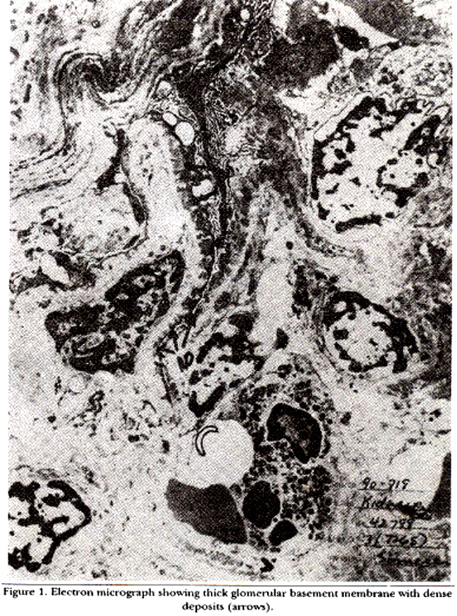

revealed irregularly thickened basement membrane with dense deposit distributed along the subepithelial aspects; these were separated and occasionally surrounded by basement membrane extensions. Epithelial foot processes demonstrated effacement and the tubules showed mild degenerative change (Figure 3).
DISCUSSION
Apart from transplant glomerulonephropathies recurrence of glomerulonephritis in a renal allograft occur in 6-9% of cases4 and these have different morphological forms like diffuse proliferative, crescentric, membranous and focal sclerosis5-7. The most common gI omerular lesion which occurs denovo is membranous GN (MGN) the reported incidence in the West being 2-9%3. MGN may occur between four months to six years following transplant with a mean Of 21 months8. The diagnosis is suspected when nephrotic syndrome is not associated with obvious clinical features of rejection, while renal function is either normal or slightly decreased. It is confirmed by excluding recurrence of GN and by the histological picture. The denovo MGN progress to renal failure more rapidly than the idiopathic form3 and the graft survival is poor9. There is no relationship with the type of graft, age, sex, race and antigen matching. To the best of our knowledge this is the first case of denovo membranous glomerulonephropathy in a renal allograft reported from Pakistan.
ACKNOWLEDGEMENT
We are extremely thankful to Prof S.A.J. Naqvi, Nephrologist, Jinnah Postgraduate Medical Centre for his advice and opinion. We are also thankful to Mr. Sulaiman his help in preparing this manuscript.
REFERENCES
1. Calne, R.Y. The currentstatua of renal transplantation. Kidney Int., 1986;30:23s-24s.
2. Brenner, D.M. and Lazarus, M.J. Chronic renal failure in Harrison’s principles of internal medicine. 12th ed New York, McGraw-Hill, 1991; pp. 1150-57.
3. Laun, T., Janice, 0., Vivette, D.A. et al. De novo membranous glomerulonephropathy in renal allograft: a report of ten cases and review of literature. Am.J.Med. Dis., 1989;2:131-44.
4. Morzycka, M., Croker, B.P. Jr., Seigler, H.P. and Tiaher, CC. Evaluation of recurrent glomerulonephritia in kidney allografts. Am.J.Med., 1982;72:588-98.
5. Camaroon, J.S. Glomerulonephritia in renal tranaplanta. Tranaplantation, 1982;34:237-45.
6. Habib, It, Antignac, C., Hinglais, N., Gagnadous, M.F. and Broyer, M. Glomerular leaiona in the tranaplanted kidney in children. Am.J.Kidney Dia., 1987;10:198-207.
7. Mathew, T.H. Recurrenceofdiaeaae followingrenal tranaplantation. Am.J.Kidney. Dis., 1988;1285-96.
8. Jag, M.D. Recurrence of glomerulonephritia following renal transplantation (Fart 1). Saudi Med. J., 1990; 11:444-49.
9. Cosyns, J.P., Firaon, Y., Squifflet, S.F., Alexandre, OF., Van Ypersele de Strihou, C, Finn, V.W., Sweet, Si., Shapiro, K.S., Cho, S. and Harrington, J.T. De novo membranous nephropathy in human renal allografts; reporta of nine patients. Kidney Int., 1982;22:177-83.
Journal of the Pakistan Medical Association has agreed to receive and publish manuscripts in accordance with the principles of the following committees: