

Abdul Ghafoor Qamar ( Department of Dermatology, Quaid-i-Azam Medical College, and B.V. Hospital, Bahawalpur. )
June 1992, Volume 43, Issue 6
Original Article

ABSTRACT
This is a pilot study of 5 nail specimens to evaluate the role of candida species in onychomycosis by taking pure isolates of different species of candida, growing these yeasts with normal nail keratin and assessing the growth of fungus periodically macroscopically and the final evaluation was made under electron microscope. The results suggest that the candida albicans primarily has an important role in keratolysis of nails (JPMA42:143, 1992).
INTRODUCTION
Onychomycosis is the invasion of the healthy nail plate by species of dermatophytes, as well as variety of non-dermatophytes (yeasts and moulds) which may cause nail infection particularly after tissue damage by trauma or disease1. Fungus infection of the nails are extremely common2 but dermatophyte onychomycosis in children is very uncommon3. Dermatophyte isolation in approximately 25% but nondermatophyte fungi in 50% of toe nails and yeasts from 25% of the nails in combination with other organisms have been reported earlier4. Candida albicans and C. parapsilosis are most commonly isolated yeasts from abnormal toe nails but C parapsilosis is the most prevalent of the two5. The role of the candida species in the pathogenesis of nail disease is complex. Onychomycosis due to candida species is thought to be restricted to patient with chronic mucocutaneous candidosis (CMC) or as secondary invaders in chronic paronychia6. The significance of candida in the pathogenesis of nail dystrophy ought to be reconsidered as some patients with some forms of nail dystrophy not associated with either mycocutaneous candidosis or chronic paronychia from which this yeast is isolated, are cured by an oral antifungal drug alone. This suggests that candida may play an important role in the development of nail dystrophy and candida species appears to be a significant pathogen in some patients with primary onycholysis of the finger nails particularly where there is underlying peripheral vascular disease or Cushing\\\'s syndrome. The aim of the present study was to evaluate the role of candida species in onychomycosis since this yeast is not considered to be either keratinolytic nor can calonise healthy nails7.
MATERIALS AND METHODS
Pure isolates of candida albicans, parapsilosis and scopulariopsis brevicaulis were maintained on glucose peptone agar. Keratinous tissue from patients who presented with suspected finger or toe nail dystrophy due to dermatophytosis but who were found to be negative on direct microscopy and culture were used in the study. Small pieces of nail (2-3 mm in dimensions) were prepared by paring larger fragments of nails with a scalpel blade. Potassium hydroxide 20% (KOH) was added to nail fragments and left for varying periods of time and direct microscopy techniques were used with light microscope for assessing the growth of yeasts on the nail fragments. Finally transmission electron microscopy was carried out in the electron microscopy unit, Department of Dermatology and scanning electron microscopy was performed in the Department of Ophthalmology, University of Glasgow/Western Infirmary.
Experiment 1
Cell lawns of each isolate were prepared on glucose peptone agar. At the same time nail fragments were placed on inoculated surface of the agar plates. The culture dishes were incubated at 37°C for one week. The growth of each fungus was assessed macroscopically and microscopically by the KOH technique after 48 hours and one week incubation to determine the penetration of fungal hyphae into the nail keratin.
Experiment 2
After 2 weeks incubation of similar surface cultures, fragments of nail were examined by direct microscopy to assess the degree of keratinolysis.
Experiment 3
Organisms were inoculated into 0.5 ml volumes of horse serum containingsmall fragments of nail tissue and incubated at 37°C. After 24 hours one nail fragment from each culture was removed and examined by direct microscopy for hyphal formation and keratinolysis. Other pieces of nail fragment were removed and processed for electron microscopy.
Experiment 4
Experiment No.3 was repeated but this time only candida albi cans was grown in horse serum containing pieces of nail to confirm the electron microscopy findings.
RESULTS
The results of experiment No.1, 2, 3 and 4 are given in
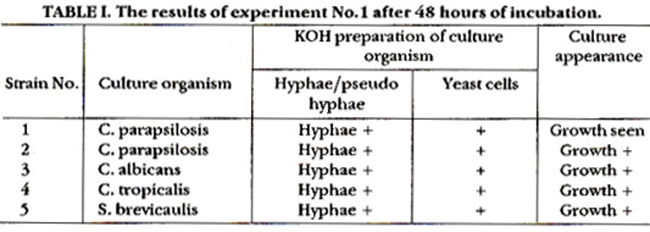
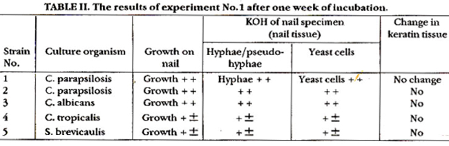
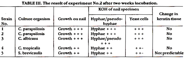
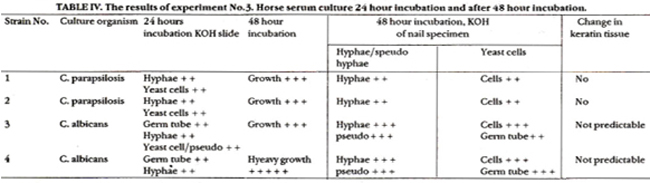
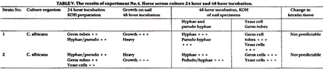
Tables I, II, III, IV and V and the electron microscopy appearance is shown in Figures 1, 2 and 3.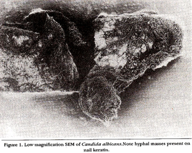
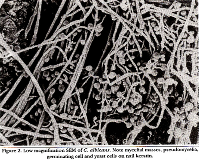
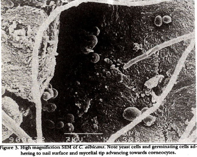
The results of experiment No.1 and 2 (Table I, II and III) show that C. parapsilosis, C. tropicolis, £ brevicanlis and C. albi cans can grow on the nail surface but no change in the nail tissue was observed. Discarding C. tropicolis and £ brevicanlis experiment No.3 was done using horse serum as nutrient medium with small fragments of nail tissue. The results of which are given in Table IV, indicate that germination by C. albicans when grown in nutrient medium-horse serum, some lysis of the nail tissue was seen under the light microscope but it was not certain that lysis was due to C. albi cans invasion or by the action of potassium hydroxide. As there was no germination by C parapsilosis so discarding C parapsilosis experiment No.4 was performed as a repeat of experiment No.3 by growing C albicans in horse serum with the nail specimens following the encouraging scanning electron microscopic results. The result of experiment No.4 is given in Table V which reconfirms the previous finding that there is some invasion of the keratinous substrate. Scanning electron microscopy (SEM) was done and recorded to visualise the nail invasion by C albicans. The scanning electron micrographs (SEM) under low magnification (Figure 1 and 2) indicated that there was heavy growth of mycelia, yeast cells, pseudomycelium and germs tubes of C albi cans on nail keratin. High magnification SEM (Figure 3) clearly showed the adherence of germinating yeast cells on the keratinous substratum and the hyphal tip advancing to invade the cellular structure of nail keratin.
DISCUSSION
Candida onychomycosis presents formidable problems in therapy8 but recently a complete clinical and mycological remission in six patients with nail infections due to C albicans not associated with paronychia, treated with itraconazole 100 mg daily for about six months has been reported9. This suggests that C albicans can invade any form of keratinised substratum in vitro but Kapica and Blank10 found that this fungus could utilize hoof and nail keratin in the presence of 2% glucose. C. albicans is the only yeast so far reported which is capable of invading the nail plate causing total dystrophy and producing a clinical picture similar to dermatophyteonychomycosis but the invasion of the nail plate is rare5. Nail plate invasion by the dermatophytes usually occurs through fronds and borers either from the lateral nail fold by forming net work of channels and lacunae leading to opacity and eventually destruction and crumbling of the nail plate forming the free edge by colonization at subungual space by producing an enzyme system. Invasion of the nail by non-dermatophytes (yeast and moulds) is simply by their presence of hyphae or yeast cells in nail material on direct microscopy and by re-isolation of the same organism on multiple occasion but S. brevicaulis can be demonstrated by electron microscopy inside the keratinized cells as reported by Acten et al12. The results of experiment Nos. 1 and 2 (Table I, II and III) indicate that C albi cans can grow on nail surface but no lysis of nail tissue was observed, but, when nutrient medium-horse serum was used to perform experiment No.3 some lysis of nail keratinwas seen under light microscopy (Table IV). The results of experiment No.4 (Table V) reconfirm the previous finding that C albicans can invade the nail keratin. Low magnification SEM showed that there was heavy growth of mycelia and yeast cells pseudomycelia and germs tubes of C albi cans on nail keratin (Figure 1 and 2). High magnification SEM clearly showed the adherence of germinating yeast cells on the keratinous substratum and the hyphal tip advancing to invade the cellular structure of nail keratin (Figure 3), similar to that as described by Pfister et al13, who reported the stages in the course of development of mycosis of the nail by SEM and found the mycelial tip going to pierce the matrix of the nail, keratinolysing its cellular structure and finally destruction of the entire nail. In the present study the onycholysis caused by the advancing hyphal tip of C albi cans cannot be predicted with certainty although it is beyond doubt that there is adherence of germinating yeast cells to the surface of nail corneocytes by their usual mode of stickiness. One can object that there is no actual growth of C albi cans in the nail matrix and this yeast has not utilized keratin as its nutrient to germinate because C. albi cans was grown in horse serum but anyhow this was in vitro study and C albicans may use nail keratin for its nutrition on the finger and toe nail in vivo. in conclusion this study confirms that C albicans is a primary pathogen of the human nail and suggests that further studies should be done to signify the important role of C albi cans as a keratinolytic yeast.
REFERENCES
1. clayton, Y.M. and Midgely, 0. Identillcation ofagents ofsuperticial mycoaes, in medical mycology: a practical approach. Edited by E.G.V. Evans and M.D. Richardaon. Oxford, Oxford University Press, 1989, pp.65-95.
2. Samman, PD. Fungua infection (tines unguium), in the nails disease. Edited by P.O. Samman. London, W.H. Medical Booka, 1965, p.39.
3. Philpot, CM. and Shuttleworth, D. Dermatophyte oxychomycosis in children. Clin. Exper. Dermatol., 1989;14:203-5.
4. Zaias, N., Oertal, I. and Elliot, D.F. Fungi in toe nails.J. Invest Dermatol., 1969;53:140-42.
5. Zaias,N. Onychomycosis. Arch. Dermatol., 1972;105:263-74.
6. Hay, R.J., Baran, R. Moore, M.K. and Wilkinson, J.D. Candida onychomycosis. An evaluation of the role of candida species in nail disease. Br.J. Dermatol., 1988;118:47-58.
7. English, M.P. Nails and fungi. Br.J. Dermatol., 1976;94:697-701.
8. Hay, R.J. and Clayton, Y.M. The treatment of patients with chronic mucocutaneous candidoaia and candids onychomycosis with ketaconazole. Clin. Exper. Dermatot, 1982;7:155-62.
9. Hay, R.J., Clayton, Y.M., Moore, M.K. and Midgely, 0. An evaluation ofitraconazolein the managementof onychomycosis. Br.j. DermatoL, 1988;119:359-66.
10. Kapica, Land Blank, F. Growthofcandida albicana on keratin assolesourceof nitrogen. Dermatologica, 19S7;11S:81-10S.
11. English, M.P. The saprophytic growth of non-keratinophilic fungi on teratinized substrata and a comparison with keratinophilic fungi. Trans. Br. Mycological Soc., 1965;48:219-35.
12. Plister-Wartha, A. and Plister, T.H. Stages in the development of mycosis of the nail as seen in the scanningelectron microscope. Fortschr. Med., 1988;106:53-55.
Journal of the Pakistan Medical Association has agreed to receive and publish manuscripts in accordance with the principles of the following committees:


