

Abrar Ahmad Javed ( Departments of Radiotherapy, Nishtar Medical College and Hospital, Multan. )
Akhtar Ali Tahir ( Departments of Surgery, Nishtar Medical College and Hospital, Multan. )
Durr-e-Sabih ( Atomic Energy Medical Centre, Nishtar Medical College and Hospital, Multan. )
Baqa Jillani ( Departments of Radiotherapy, Nishtar Medical College and Hospital, Multan. )
October 1993, Volume 43, Issue 10
Case Reports

Introduction
Cavernous haemangioma (CHL) is the commonest benign tumour of the liver. It was first described in 1861 by Frerichs1 and later by others2-5. CHL of the liver are common in females and can occur at any age. The majority of these tumours are small, asymptomatic and without clinical significance. Large tumours might cause symptoms and require treatment. The symptoms can be non-specific and diagnosis difficult. We are describing two patients of CHL whom we saw between March 1982 and 1989.
Case 1
A 36 year old (gravida 13, para 6) female was admitted in our unit in June, 1986 with a biopsy proven cavernous haemangioma of the liver. Prior to surgery she complained of having an abdominal mass for about two years. The mass had become painful 3 months before she presented for surgery. Onlaparotomy, a large (10x15 cm) vascular tumour was seen in the central region of the liver, this was considered unresectable and only a biopsy was taken confirming the diagnosis of CHL. On presentation to the radiotherapy OPD she was noted to be well built and well-nourished. Her blood pressure and pulse were normal. On examination a mass measuring 10x17 cm was visible and palpable on the right side of the abdomen extending 10 cm below the costal margin. It was soft and non-tender, the operation scar was healed. Routine haematology and liver biochemistry tests were normal. Barium meal showed a displaced stomach (Figure A).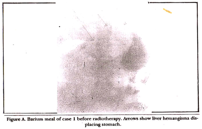
Liver scan with Tc-99m tin-colloid showed a large cold area in the central part of the liver (Figure 1).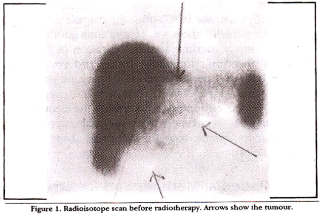
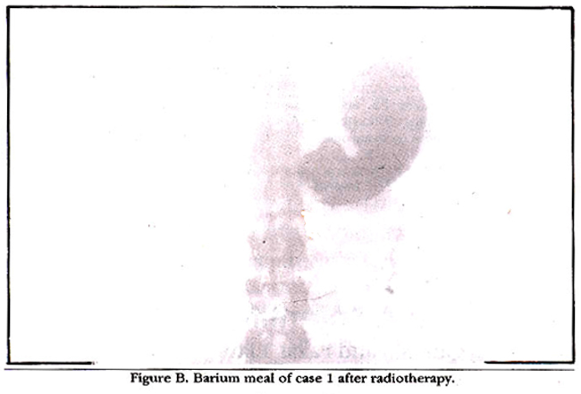
On ultrasound a complex mass with both solid and cystic elements was seen in the liver. The patient was given external radiotherapy with cobalt 60 teletherapy unit at 65 cm SSD (source skin distance), a total tumour dose of 3000 CGy (centigray)[1 CGy = 1 RadI was given five days a week over 28 days, through a 19x19 cm anterior portal designated on the basis of isotope scan and upper GI x-ray findings. The dose was calculated at the midpoint of tumour from skin (5cm). The tumour started regressing gradually 3 weeks after radiotherapy. By December, 1988, i.e., 18 months after radiotherapy, no palpable tumour remained in the abdomen. Liver scan in September, 1990 showed almost complete regression of the tumour (Figures 2 and B).
Case 2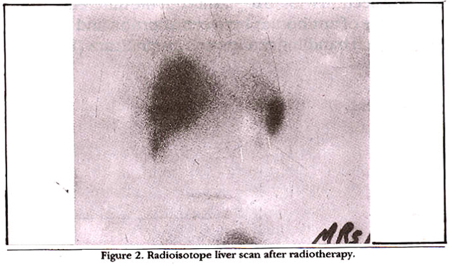
This 45 year old (gravida 13, para 11) female presented to the radiotherapy OPD on 29/3/ 1987. She had had laparotomy 2 weeks prior, to our consultation. Laparotomy was done for dysphagia with epigastric pain and mass formation. A large tumour occupying the whole left lobe was found on surgery. On admission she appeared fairly well nourished and well built Her pulse and blood pressure were normal. On examination, the operation scar was healing well, a mass was palpable below the right costal margin, extending medially into the epigastrium. There was no as cites, jaundice, peripheral oedema or fever. There was no history of nausea or vomiting though the patient did complain of dyspepsia. Investigation before surgery showed an 8x3 cm mass in the liver on ultrasound. Gastroscopy showed mild duodenitis. Investigations before radiotherapy showed normal blood counts and a negative Casoni’s test. Tests of liver function were normal except for a raised serum alkaline phosphatase (524 u/l, normal upto 11 u/l). Radiological work-up before radiotherapy revealed a cold area in the left lobe of the liver on isotope scanning. Isotope blood pool study of the liver done two days later showed gradual filling up of the cold area in liver with labelled RBCs. Ultrasound examination revealed a complex mass in the left lobe of the liver, 3.5 cm from the skin. The midpoint of the tumour was 6cm from the skin. The patient was given external radiotherapy with Cobalt-60 teleradiotherapy equipment. A total tumour dose of 1200 CGy (Centigray) at the midpoint of the tumour was delivered by an anterior portal. A daily fraction of 150 CGy five times a week using a field size of lixil cm designated according to ultrasound and isotope scan was used. The patient tolerated the radiotherapy well and she only had mild radiation sickness during the treatment. Her path disappeared starting 4 weeks after radiotherapy. There was only a slight regression of the palpable mass in the epigasthum. Ultrasound examination done 4 months after radiotherapy showed only a slight regression. Repeat isotope scan and ultrasound at 1 monthly intervals during the last one year showed no further change in the liver mass but the patient remains symptom free.
Discussion
CHL carries a risk of spontaneous haemorrhage; there is disagreement about the magnitude of this risk, but literature from Japan and USA report that this complication is fairly common6. The main treatment modalities are usually surgery and radiotherapy7. Surgery is the treatment of choice for localized CHL7,8. This should be attempted in specialized centres with experience in tumour surgery7. Radiotherapy for CHL was first used by Ray9 in 1939. A high response rate has been reported by others10-12 but because of the rarity of the disease, an optimal schedule for radiotherapy has not been fully established. The two patients reported were treated with different doses of radiotherapy. Patient 1, who had a larger tumour was treated with 3000 CGy similar to the regime of Issa and Ozakki11,14. Patient 2 was given a smaller total dose but a larger daily fraction of 200 CGy as recommended by Park and Philips15. Very good response was seen in the first patient who bad complete regression of the palpable tumour and relief of symptoms. In the second patient, there was only a slight regression of the tumour, starting 4 months after radiotherapy but her symptoms of pain and discomfort in the epigastrium disappeared. Radiation to the liver can cause hepatitis which is dose related. 3000 CGy to the whole liver is the threshold beyond which this damage can occur12. Since this dose was not exceeded we have not observed any hepatitis in our patients. Both patients are alive and symptom free after 6-1/2 and 3-1/2 years respectively. The success of treatment in this condition should be assessed by relief of symptoms and arrest of growth13,14. Radiotherapy is able to achieve this in a great majority of patients with CHL. Considering the fact that the majority of large haemangiomas are unresectable, we conclude by proposing that radiotherapy is the treatment of choice for large symptomatic cavernous baemangioma of liver.
References
1. Frerichs, F.T.A. clinical treatise on diaesse of the liver. Translated by C. Murecbison. Vol. 2. London, NewSyedenbam Society, 1861.
2. Shumaker, H.B. Haemangioma of the liver; discussion of aymptomslogy and report of patienta treated byoperation. Surgery, 1942;11:209-22.
3. Wilson, H. and Tyson, W.T. Massive bemangiomas of the liver. Ann. Surg., 1952;135:765-74.
4. Henaon, S.W., Gray, H.K. and Dockerty, M.B. Benign tumoura of the liver. Surg. Gynaecol. Obst., 1956;103:23-30.
5. Adam, Y.G., Huvos, A.G. andFortner,J.G. Gianthemangiomasof theliver. Ann.Surg., 1970;172:239-45.
6. Yaamamoto, T., Kaworda, Y., Yano, T. et al. Spontaneous rupture of baemangioma of liver. Treatmentwith transcatheter hepatic arterial embolization. AmJ.Gaatroenterol., 1991;11:1645-49.
7. Feunell, RH., Iwataucki, S. and Kano, T. et al Excisional treatment of cavernous haemangioma of the liver. Ann. Surg., 1980;192:25-27.
8. Viahrevslcy, V.A., Mohan, V.S., Pomelor, V.5. et al. Surgical treatment of giant cavernous hemangioma of the liver. HPB (hepato pancreato biliary). Surg., 1991;4:697&
9. Ray, B.S. Large cavernous haemangioma of the liver. Report of an inoperable case treated with roentgenotherapy. Ann. Surg., 1939;109:373-82.
10. Nagar, R.C. and Rhargava, K.N. Massive haemangioms of the liver.). Indian Med. Assoc., 1983;80:179.
11. Okazaki, 0., Yoshino, M., Yoshids, T. et aL Radiotherapy of hemangioma cavernosum oftheliver. Gastroenterology, 1977;73:353-56.
12. Reed, G.B. and Cox, A.J. The human liver after radiation injury. A form of veno-occlusive disease. Am.J. Pathol., 1966;48:597-607.
13. McKay, M.J., Carr, P.3. and Langlands, A.D. Treatment of hepatic cavernous haemangioma with radiation therapy. Aust. N.ZJ. Surg., 1989;12:965-68.
14. Issa, P. Cavernous haemangioma of the liver. The role of radiotherapy. Br.J. Radiol. 1968;41:26-32.
15. Park,W.C., Phillips, P. The role of radiotherapy in the management of haemangioma of the liver. JAMA., 1970;212:1496-98.
Journal of the Pakistan Medical Association has agreed to receive and publish manuscripts in accordance with the principles of the following committees:


