

Tabir S. Shamsi ( Department of Haernatology, United Medical and Dental School, Guy’s Hospital, London. )
Khalid Z. Hashmi ( SE1 9RT and Liaquat National Hospital, Karachi. )
June 1994, Volume 44, Issue 6
Review Articles

Introduction
Hairy cell leukaemia is a chronic B cell disorder which runs an an indolent but progressive course characterized by cytopenia, recurrent infections and splenomegaly1-3. The hallmark of this disease is the presence of cells with filiform cytoplasmic surface projections in peripheral blood and/or bone marrow. In recent years the introduction of new chemotherapeutic agents has revolutionized the treatment and prognosis of this condition, Interferon alpha is useful in controlling the disease, while drugs affecting nucleotide metabolism offer the possibility of lasting remission.
Epidemiology
Hairy cell leukaemia accounts for 2% of all leukaemias with an overall incidence of 2 cases per million of the population1. The median age at diagnosis is 52 years and more than half of the cases present in the fifth and sixth decade of life. Affected males outnumber females 6:1. Before the introduction of systemic therapy the median survival was 4 years although a few patients survived for as long as 25 years2-4. It remains to be seen how far new therapeutic regimens will alter the median survival in this disease.
Aetiology
The aetiology of hairy cell leukaemia is unknown5. Infection with Epstein Barr Virus (EBV), cigarette smoking, chronic anaemia, woodworking and framing, routine use of aspirin or tranquilizers and prior exposure to chemicals have all been suggested as possible predisposing factors; but no firm evidence has emerged to support any association with hairy cell leukaemia6 . Chang et al. used a genomic EBV internal repeat probe and an oligonucleotide probe directed against the EBER1 gene, but were unable to demonstrate a link between EBV and hairy cell leukaemia7. Genetic factors may be involved in the aetiopathology, since hairy cell leukaemia has been reported in patient’s first degree male relatives and to date atleast 15 such cases have been described in the literature8.
Pathology
Hairy cells infiltrate bone marrow (Figure 1),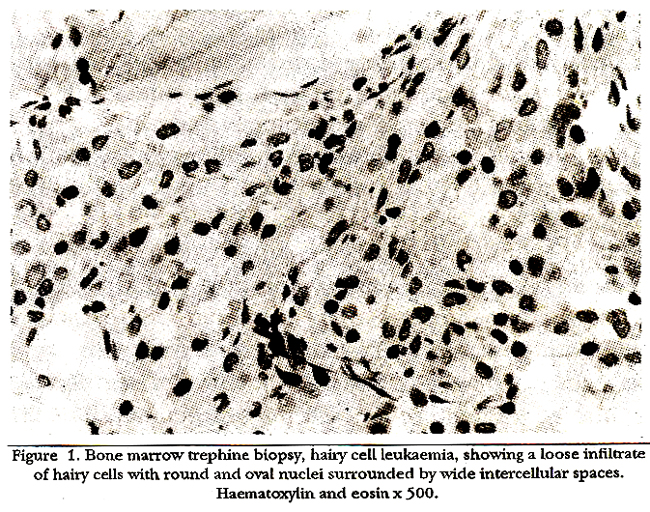
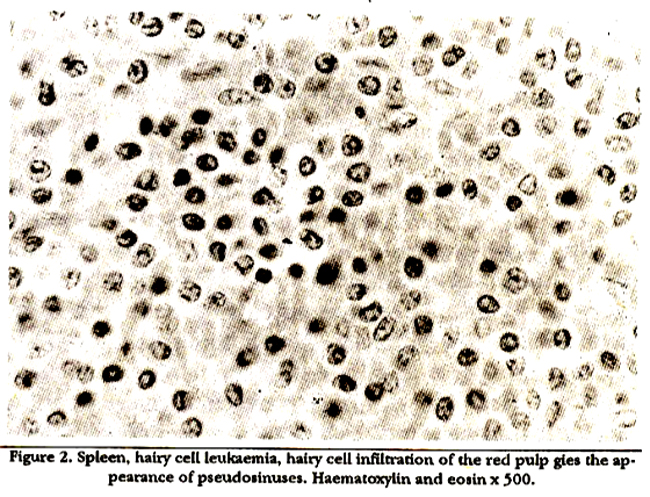
spleen (Figure 2), lymph nodes and skin. Cytopenia and organomegaly reflect the degree of organ involvement. The bone marrow shows diffuse infiltration by hairy cells, with increased fibroblastic activity and reticulin deposition; in consequence simple marrow aspiration results in a ‘dry tap’ in 40% of patients9. When marrow fragments are obtained smears typically show large mononuclear cells with abundant cytoplasm and delicate surface projections. Their nuclei are round, oval or indented with no visible nucleoli. Trephine biopsy of the marrow is advisable in all cases to support the diagnosis and to determine the response to treatment10. Histologically the marrow is replaced by a loose mononuclear cell infiltrate which may be patchy or diffuse10-13. Wide intercellular spaces give the appearance of clear zones between the abnormal cells. Connective tissue stains reveal fibrosis with increased reticulin deposition which typicallyhas a dense mesh like appearance. Electron microscopy of hairy cells shows a characteristic abnormality known as a ribosome-lamella complex in 30-50% of cells. These structures consist of a sheath of parallel lamellac with rows of evenly spaced ribosomes in between them. The spleen shows, uniform smooth enlargement in 80% of cases and histologic examination shows hairy cell infiltration of the red pulp. White pulp is scanty and pseudosinuses are formed in the red pulp resultingin the appearance of so called red cell lakes14. Lymphadenopathyis rare in the early stage of the disease but when present is associated with a diffuse inifitrate of widely spaced hairy cells3.
Clinical Features
Most patients seek medical attention on account of non-specific symptoms of ill health such as weakness, weight loss, tiredness, lethargy and shortness of breath, infection of the consequences of cytopenias5. Pyogenic infections with gram positive Cocci and gram negative bacilli are common in neutropenic patients, but infections with atypical mycobacteria and toxoplasmosis also occur frequently suggesting defective cell mediated immunity. Bone pain, vasculitic rash, arthritis, polyarteritis nodosa and systemic vasculitis may complicate the course of the disease at some stage but occasionally are present at the time of diagnosis. Splenomegaly is detectable by clinical examination in approximately 80% of patients; the spleen is more than 10 cm below the costal margin in about a quarter to one third of them2,5. There is hepatomegaly in 30-40% of cases5. Lymphadenopathy was recently reported in 8 out of 27 new patients (30%) in whom computerized tomographic scans (CT scan) of the abdomen were performed at diagnosis and in 19 out of 50 patients 08%) scanned during the course of their disease, but clinically apparent lymph node enlargement is unusual. Lymphadenopathyis associatedwith significant splenomegaly, long standing disease and resistance to treatment15.
Laboratory Features
At presentation patients with hairy cell leukaemia as a rule show one or more cytopenias or even pancytopenia depending upon the extent of marrow infiltration and splenomegaly. The total white cell count maybe low, normal, or high, but is rarely more than 20x109/12,5. Neutropenia and monocytopenia are usual9,16. Hairy cells are found in the blood in up to two thirds of cases (Figure 3).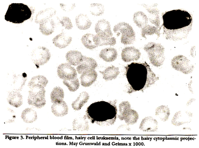
A minority have few or no detectable hairy cells on blood film examination. Mild to moderate thrombocytopenia is found in 80% of cases, but symptoms due to severe thrombocytopenia are infrequent. Red cells are often mildly macrocytic and show anisocytosis9. Anaemia is usually the result of bone marrow infiltration and replacement by malignant cells2,5. Hairy cells produce a factor or factors, possibly tumour necrosis factor (TNF) which inhibits granulocyte/macrophage colony forming units (CPUGM) and erythroid burst forming units (BFU-E) in vitro and which along with other as yet unidentified factors may play a part in haemopoietic inhibition18-20. There is usually an increase in plasma alanine transaminase and gamma glutamyl transferase, possibly caused by hairy cell infiltration of hepatic sinuses and portal traéts. A positive reaction for isoenzyme \\\'5 of acid phosphatase which is resistant to treatment with tartrate (TRAP yositive) is a characteristic feature of hairy cells3,17. Only a small minority of cells in other lymphoproliferative disorders may give a positive TRAP reaction17. The neutrophil alkaline phosphatase score is elevated in patients who have a normal neutrophil count. Hairy cells showfeatures of activated B lymphocytes21,22. They also express aberrant immunologic markers of monocytes as well as activated T lymphocytes. Immunophenotyping shows strong surface membrane IgA and IgG expression and positive reactions with monoclonal antibodies to CD 19, CD2O and CD22, which are B cell markers. Positive reactions with FMC7, HC2, CD25 and CD11c antibodies are also usual4,22. It is likely that atleast 25% of cases of hairy cell leukaemia show clonal chromosomal abnormalities; chromosomes 14 and 12 are involved in the majority of these, but at present the breakpoints and association with oncogenes are not known24-27.
Diagnostic Criteria
Characteristic hairy cells seen on blood film examination associated with splenomegaly are virtually diagnostic28. Trephine marrow biopsy showing typical histological changes and the demonstration of TRAP positive cells in blood or marrow together with appropriate immunologic cell surface markers confirm the diagnosis. Difficulty may arise when few or no hairy cells are found in the peripheral blood The clinical conditions which should be considered in the differential diagnosis are CLL with isolated splenomegaly28 splenic lymphoma with villous lymphocytes23 prolymphocytic leukaemia, myelofibrosis and aplastic anaemia9,28. If few or no hairy cells are seen on peripheral blood examination, marrow aspirate yields only a dry tap and the trephine biopsy is. hypoceilular, aplastic anaemiamay be mistakenly diagnosed The clinical findings and the results of the examination of the patient’s blood film and bone marrow supported by the demontration of appropriate cytochemical and immunologic markers establish the diagnosis in almost every case.
Treatment
The treatment of hairy cell leukaemia has changed dramatically in the last ten years. With the advent of effective chemotherapy, the aim of treatment has shifted from palliation to eradication of the disease. Until recently, patients with minimal or no symptoms were often left untreated. Symptomatic patients were frequently splenectomized at some stage of the disease and received supportive treatment including blood transfusion and antibiotics as necessary. Systemic treatment for hairy cell leukaemia should if possible be started well before the development of profound cytopenia or other maj or complications.
Splenectomy:
Until recently splenectomy was considered the treatment of choice for hairy cell leukaemia29,30. In up to 50% of patients, blood counts return to normal after the operation, while amongst 30-40% of the remainder there is some improvement in atleast one blood cell line. Although an occasional patient enters lasting remission, the majorityrelapse and the disease gradually progresses if no other treatment is given. Presumably splenectomy removes the main site of cell sequestration as well as removing a major part of the tumour bulk31,32. It is sometimes followed by complete reversal of marrow findings, an effect which is difficult to explain. The response to splenectomy may be rapid, the platelet count recovering within two weeks of operation followed by increases in neutrophil numbers and haemoglobin over subsequent months. Non-responders have a poor prognosis without systemic therapy31,32. Since the advent of systemic drugs the role of splenectomy has decreased, but the operation may still be considered in those who fail to respond to interferon, deoxycoformycin or chlorodeoxyadenosine. It may also be considered in patients with symptoms due to massive splenomegaly.
Cytoxic Chemotherapy:
Systemic chemotherapy with or without splenectomy was frequently employed before the introduction of interferon. Despite the occasional claims of good responses obtained with cytoxic agents including chlorambucil, cyclophosphamide, anthracyclines, steroids and vinicristine alone or in combination, any beneficial effects were as a rule minimal and transient34-37.
lnterferonAlpha:
In 1984, Quesada et al. reported seven patients treated with interferon alpha (IPN a) 3 million units/day by intramuscular injection for atleast six months. Three patients achieved complete remission and four showed partial responses. These effects were maintained for many months after stopping the drug39. Several investigators have confirmed Quesada’s observations40-48. Interferon is effective in up to 80% of patients treated, 70% achieving partial remission and 5-10% a minimal response with improvement of only one blood cell type. Only a minority of patients (5%) enter complete remission46 . The type of alpha interferon used, whether the product of a cultured lymphoblastoid cell line or a recombinant DNA product appears to make little difference in the response to treatment. In patients who enter complete remission, the platelet count returns to normal within approximately two months of starting treatment. Haemoglobin may take four months and neutrophils four to six months to recover39-41. Hairy cells decrease in the peripheral blood within four weeks of therapy. These improvements in peripheral blood count are the direct result of a gradual reduction in bone marrow infiltration accompanied by an expansion in haemopoietic tissue. Complete eradication of hairy cells from the marrow is rare. Marrow fibrosis usually persists despite long term treatment with interferon. The optimal dose and duration of treatment is not well defined40-48. The dose most commonly used for recombinant interferon alpha 2b (IFN alpha 2b) is 3 million units 3 times a week by subcutaneous injection for 12-18 months while for interferon alpha 2a (IFN alpha 2a) it is 3 million units daily by subcutaneous injections for the same duration. Low dose IFN 0.2 million units per square meter of body surface area (BSA) per day is less toxic but also less effective than “standard dose” IFN alpha49. Higher doses are less well tolerated. A recent trial evaluated the duration of IFN treatment in two prospective groups48. One group received the drug for 12 months and the other for 18 months. This study did not find significant differences in peripheral blood count in the two groups at the end of IFN treatment. The number of patients requiring retreatment in both groups was similar. The mechanism of action of IFN alpha is not fully clear. Its direct effects on hairy cells include modification of gene expression, induction of specific protein synthesis and modulation of cytokine production. These actions induce hairy cells to differentiate and make the cells resistant to growth factor stimulation. Interferon alpha also activates the production of natural killer (NK) cells50.Interferon alpha is usually well tolerated at the “standard dose48. During the first week or so, many patients feel influenza like symptoms which may include fever, chills, headache, malaise, myalgia and tachycardia. Paracetamol 1 gram half an hour prior to each injection ameliorates these symptoms. It is often recommended that interferon should be injected at night so that the side effects are not as troublesome. On continued treatment these iatrogenic symptoms usually resolve. Myelosuppression is a frequent side effect and a proportion of patients receiving prolonged treatment complain of chronic fatigue48. Other rarer side effects include dry skin, diarrhoea, constipation, depression, paraesthesia and loss of libido. Up to 30% of patients treated with IFN alpha 2ahave been found to develop neutralizing antibodies51. These antibodies disappear on long term follow up despite continued 1PM alpha 2a injections51. Neutralising antibodies are rarely formed against 1PM alpha 2b52,23. In cases where antibodies cause refractoriness to IFN alpha 2a, TEN alpha 2b can be used without cross reactivity; alternatively, treatment with deoxycoformycin or deoxyadenosine may be considered.
Deoxycoformycin (Pentostatin):
Deoxycoformycin (2-DCP) is an inhibitor of adenosine deaminase (ADA), an enzyme which is necessaw for lymphocyte proliferation and functions. The exact mode of action of this thug against hairy cell leukaemia is not certain but there is evidence that lympho-cytotoxicity is mediated by the accumulation of deoxyadenosine triphosghate leading to ribonucleotide reductase inhibition54-56. . Breakage of single stranded DNA, depletion of NAD and inhibition of intracellular methylation may be produced by 2-DCf56. Because of the high adenosine deaminase levels in lymphoid tissue, 2-DCE is active against hairy cell leukaemia and other lymphoproliferative disorders. Spiers et al. reported 2-DCP activity in hairy cell leukaemia in 2 patients and since then other groups of investigators have confirmed a high degree of activity against this disease54-56. The overall complete response rate is 50-80%29. The response is quicker than to IPN and peripheral blood counts start to improve within two weeks of starting treatment. 2-DCP is active in both untreated and previously treated patients57,58. The recommended dose is 4 mg per square meter of body surface area every 2 weeks for 8-10 cycles or until remission is achieved, followed by 2 more cycles as consolidation. Grever et al.59 reported the results of a randomized trial comparing 2-DCP (4 mg per square meter of BSA every 2 weeks) with IFN alpha (3 million units 3 times a week subcutaneously) in 356 untreated patients. Of the patients in the 1PM arm, 11% achieved complete remission and 38% partial remission. In the 2-DCP arm 69% entered complete remission (CR) and a further 6% partial remission (PR). The relapse free survival was significantly longer in the 2-DCF arm; 50% of those who achieved CR relapsed at 10-19 months in the 1PM group while only 4% in the 2-DCP group relapsed after 13-37 months follow up. Side effects54-58 of 2-DCF include nausea, vomiting, skin rashes, keratoconjunctivitis, fever, renal impairment and neutropenia. It should be used with caution in patients with renal insufficiency and acute infections. Urba et al60 have shown a fall in T lymphocytes in 13 patients treated with 2-DCP alternating with IPN. Both CD4 and CD8 count decreased to less than 200cells/microlitre in all patients throughout 14 months of treatment and 6 months thereafter. Levels of CD4 and CD8 returned to normal in only 4 cases. Localized infections with herpes zoster virus have been reported during treatment.
2-Chlorodeoxyadenosine (Clidrabine):
2-Chlorodeoxyadenosine (2-CdA) is a purine analogue in which chlorine is substituted for a hydrogen atom at position 2 of the purine ring. This substitution renders the drug resistant to adenosine deaminase61,62. As a result 2-CdA accumulates in the lymphoid tissues of the treated patients as chlorodeoxyadenosine triphosphate. The enzyme deoxycytidine kinase phosphorylates 2- CdA to chlorodeoxyadenosine triphosphate leading to DMA strand breaks in lymphocytes, decreased RNA synthesis and depleted energy production61,62,65. It has been suggested that 2-CdA induces apoptosis (programmed cell death) in sensitive cells61. Its exact mechanism of action is not fully established, but like 2-DCP, it is active against many lymphoproliferative disorders. Precinical studies in lymphoblastoid cell lines have shown that both B and T cells are sensitive to 2-CdA, but T cell lines are affected by a lower concentration of the drug66. Piro et al. 63 reported 86 patients who received single course of 2-CdA at a dose of 0.1 mg/kg/day by continuous intravenous infusion for seven days. These patients included both treated and untreated groups. Complete remission (CR) was obtained in 80%, while most of the remaining 20% achieved good partial remission (PR). Previous treatment did not affect the response rate. Tailman et al. 67 reported 26 patients treated with 2-CdA at 0.1 mg/kg/day for seven days as IV infusion; 15 of the 26 patients were previouslyuntreated while 11’had had treatment with 1PM alpha or 2-DCF or had been splenectomized. After a single course of 2-CdA, 16 of the 20 evaluable patients were in CR, while 4 obtained PR. Of the 4 patients who showed a partial response, 3 had residual disease in the marrow and received a second course of 2-CdA; 2 of these subsequently achieved CIt In summary, 18 out of 20 evaluable patients (90%) attai9d CR after 1 or 2 cycles of 2-CdA. Juliusson and Lilliemark68 and Estey et al. 69 have also reported a CR rate of 80% after a single course of 2-CdA. These studies have shown that 2 CdA has a much greater activity against hairy cell leukaemia than any other form of treatment, with sustained complete response rates after a single course approaching 80% Such results have attracted many research groups to use it as first line drug offering the possibility of cure. The major side effect of 2-CdA is a culture negative febrile episode occurring in 40% of neutropenic patients which it has been suggested may be due to lysis of hairy cells63,65,70. Apart from this drug related effect, infections with candida, cytomegalovirus and other fungal. infections have been reported as a cause of fever. 2-CdA has been reported to reduce the number of both B and T lymphocytes in the peripheral blood. CD4 and CD8 counts drop and may remain low for as long as 12 months after treatment64. The encouraging results of treatment with 2-CdA will require long term follow up, to determine how often CR equals permanent cure. The long term consequences of immunosuppression following its use will also need further study.
Conclusion
Large randomized studies are needed to evaluate the results of combined therapy with IFN, 2-DCF and 2-CdA compared to those obtained using a single agent. The last 10 years have seen dramatic advances in the treatment of hairy cell leukaemia which have changed therapeutic intentions from palliation to cure. It remains to be seen whether complete remissions after treatment with 2-CDF and 2-CdA amount to permanent cure in a substantial proportion of patients.
Acknowledgements
I am grateful to Dr. K.G.A. Clark, Consultant Haematologist, Guy’s Hospital, for his suggestions and comments, Col. Dr. Zahur –ur- Rehman, Consultant Haematologist, Army Medical College, Rawalpindi and Mrs. Pip Farnsworth, Secretary, Guy’s Hospital for their encouragement and secretarial assistance respectively.
References
1. Bouroncle, B.A., Wiaeman, B.K. and Doan, C.A. Leulcamic reticuloendothelioais. Blood, 1958;13:609-30.
2. Golomb, H.M., catovslcy, D. and Golde, D.W. Hairy cell leukaemia: a clinical review baaed on 71 caaea. Ann. Intern. Med., 1978;89:677-83.
3. Yam, LT., Janckila, A.J. and Lam, W.K.W. Cytochemiatry of tartrate-resistant acid phosphatase: 15 year\\\'s experience. Leukaemia, 1987;1:285-88.
4. Jansen, 3., LeBien, T. and Keraey, J.H. The phenotype ofthe neoplasticcells of hairy cell leukaemia studiedwith monoclonal antibodies. Blood, 1982;59:609-14.
5. Flandin, C., Sigaux, F., Sebahoum, C., tal. Haity-cell leukaemia: cliniealpreaentation and follow-up of 211 patients. Semin. Oncol., 1984;11(suppl2):458-71.
6. Olealce, D., Golomb, H. and Farber, M. Acase control enquicyinto the aetiology of hairy cell leulcaemia. Am.J.Epidemiol., 1985;129:675-83.
7. chang, K.L, chen, Y. and Welas, LM. Lack of evidence of Epstein- Barr virua in the hairy cell leulcaemia and monocytoid B-cell lymphoma. Hum. Pathol., 1993;24(1):58-61.
8. Gramatoici, M., Bennett, 3M., Hiscock, J.G., et al Three cases of familial hairy-cell leukaemia. Am.J. Hematol., 1993;42:337-39.
9. Cawley, J.C., Burns, G.F., Hayhoe, P.O_I. Hairy cell leukaemia. New York, SpringerVerla& 1980.
10. Burke, J.S. Thevalue of the bone-marrow biopsy in the diagnosis of haicycell leukaemia. Am.J. din. Patbol., 1978;70:876-84.
11. Bartl, R., Friach, B., Hill, W., et at Bone marrow histology in hairy cell leukaemis: identification of subtypea and their prognostic significance. Am.J.Clin.Pathol., 1983;79:531-45.
12. Lee, W.M.F. and Beckatead, J.H. Hairy cell leokaemia with bone marrow hypoplasis. Cancer, 1982;S0:2207-10.
13. Bain, B., clerk, 0. and Lampert, 1. Bone marrow pathology. Oxford, Balckwell Scientific Publications, 1992.
14. Lewis, S.M., Catovskym D., Howa, 1, et al. Splenic red cell pooling in hairy cell leulcaemia. Br.J. Haematol., 1977;35:351-57.
15. Mercieca, 3., Puge, M., Matotes, B., et at Incidence and significance of abdominal lymphadenopathy in hairy-cell leukaemia (abstract). Blood, 1993;82.2273.
16. Seshadri, R. Brown, E. and Zipursky, A. Leuksemic reticuloendotheliosia. A failure of monocyte prodoction. N.Bngl.3.Med., 1976;295:181-84.
17. Katayama, L and Yang, 3. Tartrate-reaistant acid phospbataae reactions not specific for hairy cell leokaemia. Am.J.Clin.Pathol., 1979;68:482-86.
18. Taniguchi,N., Kuratsune,H., Kanamaru,A., ecal.lnhibition against CFU-C and CFU-B colony formation by aoluble factor(s) derived from hairy cells. Blood, 1989;73:907-13.
19. Janckila, A., Gentile, P., Yam, L Haemopoietic inhibition in hairy-cell leokaemis. Am.J.aematol., 1991;38:30-39.
20. Lauria, F., Bagnara, 0., catani, L, et al. The inhibitory effect of serum from hairy cell leoksemis patients on normal progenitor cells may ditappear following prolonged treatmencwich alpha interferon. BrJ.Haemacol., 1989;72:497-501.
21. Anderson, K., Boyd, A, FIsher, 0., etal. Hairy-cell leukaemis: a tumour of pre-plasma cells. Blood, 1915;65:620-29.
22. Janaen,J., Schuit, H.R.B., Meijsr,L.M., etal. cell markers in hairy cell leukaemiaatudied in celia from 51 patients. Blood, 1982;59:52-60.
23. Neiman, R.S., Sullivan, AL and Jaffe, R. Malignant lymphoma simulating leokaemic reticoloendotheliosia: a clinicopathologic study often cases. Cancer, 1979;43:329-33.
24. Juliuaaon, 0. and Gahrton, G. Cytogenetics in CLL and related disorders. Clin-Haematol., 1993;6:821-48.
25. Brito-Babapulle, V., Pittman, S., Melo, 3., et al. The 14q +markerin hairy cell leukaemia. A cytogeneticacudy of 15 cases. Leukaemia Res., 1986;10:131-38.
26. Golomb, H., Lingren, V. and Rowley, 3. chromosome abnormalities in patients with hairy cell leukaemia. cancer. 1978;41:1374-80.
27. Golomb, H., Lingren, V. and Rowley, 3. Hairy-cell leukaemia: an analysis of the chromosomes of 26 patients. Virchows Arch (B)., 1978b;29:113-20.
28. Jansen, 3., Den Ottolander, 0., Te Velde,J. and Lopes Cardozo, P. The clinical diagnosis of hairy-cell leukaemis. Acts. Flaematol. (Bssel)., 1980;64:181-86.
29. Golomb, H.M. and Vardimsn, 1W. Response to spleneecomy in 65 patients with hairy cell leuksemis: an evaluation of spleen weight and bone marrow involvement. Blood, 1983;61:349-52.
30. Jansen, 3. and Harmans, 3.0. Spleneccomy in hairy cell leukaemia: a retrospective multicenter snalysia. Cancer, 1981;47:2066-76.
31. Jaiyesimi, L, Ksntarjian, H. and Eacey, R Advancea in the therspy for hairy cell leuksemia. Cancer, 1993;72:5-16.
32. Golde, OW. Thet-apy of hairy-cell leukaemis. N.Engl.J.Med., 1982;307:495-96.
33. Plataniss, L, Golomb, H. Hairy-cell leulcsemia. Clin.Hsemstol., 1993;6:887-98.
34. Golomb, H., Schmidt, K snd Vardiman, K Chlorsmbueil therapy of twenty four post-splenectomy patients with progressive hairycell leulcaemia. Semin. Oncol., 1984;11 (suppl 2):502-6.
35. Stewart, D.J., Benjamin, R.S., McCredie, KB., et sI. The effectiveness of rubidazone in hairy cell leuksemis (leukaemic reciculoendotbeliosis). Blood, 1979;54:298-304.
36. Mcdsrthy, 0. and Cstovsky, 0. Response to doxorubicin in hairy cell Leukaemia. Scand.J.Haemacot, 1978;21:445-47.
37. Csse, 0. Combination chemotherapy of hairy-cell Ieukaemia with cyclophosphamide, vinci-istine and prednisone. J.Msine Med. Assoc., 1980;71:36-37.
38. Cheever, M.A., Fefer, A., Greenberg, P.O., et sI. Treatment of hairy-cell leukaemia with chemo-radiotherspy and identical twin bone marrow transplantation. N.Engl.J.Med., 1982;307:47-81.
39. Quesads, JR., Rueben, 3., Manning, J.T. et al. Alpha interferon for induction of remission in hairy cell leukaemis. N.Engl.J.Med., 1984;310:15-18.
40. Wormsn, CF., Cscovslq, 0., Bryan, P.C., cc at interferon is effective in bsiry-cell leoicsemia. Br.J.Haematol., 1985;60:759- 63.
41. Qoessda, 3., Hersb, R, Manning, 3., et al. Treatment of hairy- cell leulcaemis with recombinant alpha interferon. Blood, 1986;68:493-97.
42. Lauria, F., Fos, R., Raspsdori, 0., et sI. Treatment of hairy- cell leukaemia with alpha interferon (slphs-IFN). BorJ.Cancer dIm. Oncol., 1988;24:195-200.
43. Hagberg. H., Aim, 0., Bjorlcholm, M., et sl. Alphs interferon trestmenc of patients with hairy cell leuksemia. Scand. 1 Hsematol., 1985;35:66-70.
44. Foon, K., Maluish, A., Abrsms, P., et al Recombinant leulcoryte: a interferon therapy for sdvsnced hairy-cell leulcaemis: therapeutic and immunologic results. Am.J.Med., 1986;80:351-56.
45. Golde, D., Jacobs, A., Glaspy, J., et al. Hairy-cell leuksemis: biology and treatment. Semin.Oncol., 1986;23 (suppl 1):3-9.
46. Msndelli, P., Annino, L, Cafolla A., et aL Hairy cell leukaemis: preliminary resoltswith alpha 2(r) interferon. Tumori, 1986;72:153-56.
47. Berman, B., Hellsr, G., Kempin, S., etaL Incidence of response and long term follow-up in pscientawith hsirycell leukaemia treacedwith recombinantinterferon alpha 2a.Blood,1990;75:839- 45.
46. Golomb, H., Ratain,M., Fefer, A, etal. Rsndomized studyofthe duration oftreatmentwith interferonalphs 2b in pslientswith hairy-cell leukaemia. J.Natl.Cancer.Ins., 1988;80:369-72.
49. Moormeier, 3., Ratain, M., Westbrook, C., cc at Low dose interferon slpha-2b in the treatment of hairy-cell leolcsemia. J.Natl.Cancer Inst, 1989,81:1172-74.
50. Vedantham, S., Gamlel, H-and Golomb, H.M. Mechanism of interferon action in hairycell leukaemia: a model of effective cancer biocherapy. Cancer Res., 1992;52:1056-66.
51. Steis, R.G., Smith, 3W., Urbs, W., cc sL Resistance to recombinant interferon alpha 2a in hairy-cell leuksemis associated with neutralizing anti-interferon antibodies. N.Engl.J.Med., 1988;3l8:1409-13.
52. Moormeier,1, Westbroolç C., Ratsin, M., Golomb, K Interferon alphs-2b antibodies and clinical resistance in a pstient with hairy cell leukaemia. Leuksemis snd-Lymphoma, 1989;1:43-45.
53. Spiegel, R., Spicehandler, 3., Jacobs, & and Oden, B. Lowincidenee of serom neutrslzing factors in patients receiving recombinant slpba-2b interferon (intron A). Am.J.Med., 1986;80:223-28.
54. Spies-a, A, Parekh, S. and Bishop, M. HsiLy-cell leukaemia: induction of complete remission withpentoatatin (2 deoxyeofromycin). J.Clin.Oncol., 1984;2:1336-41
55. Johnston, 1, Eisenhsuer, B., Corbett, W., ecaL Efficacy of 2. deoxycofromycin in hairycell leulcsemia: a study of the Nstionsl Cancer Institute of Canada Clinical Trials Group.J.Natl Cancer Inst 198;80:765-69.
56. Grem, J., King, S., Cheson, B. st al. Pencostacin in hairy-cell leulcsemis: treatment by special exception mecbanism. J.Natl.Cancer Inst., 1989;81:448-53.
57. Kraut, B., Bouroncle, B. and Orever, M. Pentoatatin in the treatment of advanced hairy-cell leukaemia. J.Clin.Oncol., 1989;7:168-72
58. Ho, A., Thaler, 7., Mandelli, F., et al. Reaponae to pentoatatin in hairy-cell leukaemia refractory to interferon-alpha. J.Clin.Oncol., 1989;7:1533-38.
59. Orever, M., Kopecky, K., Head, D., et al. A randomized eompariaon of deoxycofromycin (DEC) versua alpha-2a interferon (IFN) in previoualy untreated patients with hairy cell leulcaemia (HCL): an NCI-sponsored integroup study (SWOG, ECOG, CALGB, NCIC, CTG)(Ahatract). Proc.Am.Soc.Clin.Oncol., 1992;11:264.
60. Urba, W., Baseler, M., Kopp, W., et al. Deoxycofromycin induced immunosuppreaaion in patienta with hairy-cell leukaemia. Blood, 1989;73:38-46.
61. Carrera, C., Tecai, C., Piro, L., et al. 2CdA chemotherap triggera programmed cell death in normal and malignant lymphocytea (abstract). InLJ.Purine, Fyrimidine Res., 1991;1 (auppi. 1):38.
62. Seto, S, Carrera, C., Kubota, M., et al. Mechanism of deoxyadenosine and 2-chlofodeoxyadenosine toxicity to non- dividing human lymphocytes. J.Clin.Invest., 1985;75:377-83.
63. Piro, LD., Carrera, C.J., Carson, D.A., etal. Lastingremissions in hairy-cell leukaemia induced bya aingleinfusion of 2-chlorodexyadenosine.N.Engl.J.Med.,1990;322:1117-21.
64. Piro, L., Saven, A., Ellison, D., et al. Prolonged complete remission following 2-chlorodeoxyadenoaine (2-CdA) in hairy cell leukaemia (HCL)(abatract). Proc.Am.Soe.Clin.Oncol., 1992;11:259.
65. Piro, L 2-chlorodeoxyadenosine treatmentoflymphoid malignancies (editorial). Blood, 1992;79:843-45.
66. Carson, D., Waaaon, D., Kaye, 3., et at Deoxycytidine Icinase- mediated toxicity of deoxyadenosine analogs toward malignant human lymphoblasts in vitro and toward murine L1210 leukaemia in vivo. Proc.Nstl.Acad.Sci., USA., 1980;77:6865-66.
67. Tallman, M., Hackimian, D., Variakojia, D., et al. 2- chlorodexyadenoaine in the treatment of haicy cell leukaemia (abstract). Blood, 1991;78:127.
68. Juliuaaon, 0., Lilliemark, 3. Rapid recovery from cytopenia in hairy cell leukaemia after treatmentwith 2-chloro-2. deoxyadenosine (CdA): relation to opportunisticinfections. Blood, 1992;79:88&-94.
69. Estey, E., Kurzrock, R., Kantarjian, H., et al. Treatment of hairy-cell leukaemia with 2-chlorodexyadenosine (2-CdA). Blood, 1992;79:882-87.
70. Saven, A. and Pico, LTreatment of hairy-cell leukaemia. Blood, 1992;79:1111-20.
Journal of the Pakistan Medical Association has agreed to receive and publish manuscripts in accordance with the principles of the following committees:


