

Figen Kadakal ( Yedikule Chest Disease and Surgery Education and Research Hospital, Istanbul, Turkey. )
Gulfidan Aras ( Yedikule Chest Disease and Surgery Education and Research Hospital, Istanbul, Turkey. )
Dilck Kanmaz ( Yedikule Chest Disease and Surgery Education and Research Hospital, Istanbul, Turkey. )
Sevim Purisa ( Department of Biostatistics, Medical Faculty of Istanbul University, Istanbul, Turkey. )
Meltem Uzumcu ( Department of Microbiology, Taksim Education and Research Hospital, Istanbul, Turkey. )
Elvan Iatk ( Department of Internal Medicine, Haydarpash Education and Research Hospital, Istanbul, Turkey. )
Dorina Bratfalean ( Expert Biostatistician, FIRALIS SAS, Huningue, France. )
July 2013, Volume 63, Issue 7
Pilot Study

Abstract
Objectives: To see if high-sensitivity C-reactive protein levels increase even in the early stages of asthma, and to evaluate if corticosteroid therapy affects the levels in asthma patients.
Methods: The case-control pilot study was conducted at Yedikule Chest Disease and Surgery Education and Research Hospital, Turkey, from February to April 2011. Patients newly diagnosed with asthma who reported symptoms that occurred six months before diagnosis were included in the study. The protein levels were measured pre-treatment and one month post-treatment. In addition, pulmonary function test and total Immunoglobulin-E measurements were taken and the prick test was performed. Statistical analysis was done using SPSS 15.
Results: There were 15 cases; 8 (53%) females and 7 (47%) males. Besides, there were 19 Controls; 9 (47%) females and 10 (53%) males. The mean age of the Cases was 29.13±10.30 years, while for the Controls it was 28.9±5.35 years. The difference was not statistically significant (p<0.54). The difference in protein levels pre and post-treatment was not significant. However, a higher level in the pre-treatment period was found compared to the Controls. Post-treatment levels in the Cases were not significantly different than the Controls.
Conclusion: Elevated high-sensitivity C-reactive protein levels in asthmatic patients may indicate an increased risk for cardiovascular disease. Future studies in asthma patients should focus on this relationship.
Keywords: High-sensitivity C-reactive protein, Asthma, Turkey. (JPMA 63: 893; 2013).
Introduction
Asthma is characterised by airway inflammation involving various cells and mediators, including eosinophils, mast cells, T-lymphocytes, neutrophils, and cytokines.1 These agents are inducers of systemic inflammation. Additionally, increased levels of serum fibrinogen and amyloid A in asthmatics and a positive relationship with the serum acute phase reactant have been reported. Together, the data supports the existence of systemic inflammation in asthma.2,3
High-sensitivity C-reactive protein (HsCRP) is a characteristic marker of inflammatory processes and is, therefore, a good diagnostic tool for ongoing inflammation.4 Recently, it has been reported that serum hsCRP is useful for the detection of low-grade inflammation. Accordingly, hsCRP is a good maker for diseases characterised by low-level inflammation, such as cardiovascular disease (CVD) and diabetes mellitus (DM). Additionally, hsCRP levels are strongly predictive of cardiovascular events and are associated with the severity of coronary atherosclerosis.5-7 Several studies have indicated that serum hsCRP levels may be suitable for the detection of systemic inflammation in asthma.8-11 These studies showed an association between decreased pulmonary functions and increased levels of serum hsCRP, which suggests that hsCRP could be surrogate systemic marker that reflects the degree of local airway inflammation.8-11 Pulmonary function test (PFT) is the only assay of clinical reflection of airway inflammation. Corticosteroids can reduce CRP and other circulating inflammatory cytokine levels in acute pro-inflammatory states.12,13
There are two previous studies examining the degree to which corticosteroid use suppresses inflammation and leads to a decrease in CRP.14,15 In one study, the treatment and non-treatment groups were randomly selected from different patient populations.15 In the other study, patients were evaluated after three months.14
To better understand the effects of inhaled corticosteroids on hsCRP levels and to determine the diagnostic usefulness of this biomarker, the serum levels of hsCRP were longitudinally compared in the same group of patients prior to and after the corticosteroid therapy. Additionally, these parametres were compared with healthy controls. The goal of the current study was to determine the early effects of corticosteriods on asthma symptoms, especially since the disease causes systemic inflammation even in patients with only moderately severe asthma.
Patients and Methods
The case-control study was conducted from February to April 2011 at the Yedikule Chest Disease and Surgery Education and Research Hospital, Turkey. Initially, 20 patients with recently diagnosed moderate persistent asthma who had experienced symptoms for at least six months were included. The asthma diagnosis was established according to the Global Initiative for Asthma (GINA) guidelines. The features of moderate asthmatics include breathless during talking, prefer sitting, talk in phrases, usually agitated, respiratory rates increased, PEF after initial bronchodilator predicted approximatelly 60-80%, and SaO2 90-95%. All of the asthma patients were partly controlled after one month accordig to GINA version 2011.16
Besides, 19 healthy, non-smoker volunteers of similar age and body mass index (BMI) were used as the control group. The study was approved by the institutional ethics committee, and informed consent was obtained from all the participants. The study will continue in the future.
The multi-factor inclusion criterion was: patients who were recently diagnosed with moderate severity of asthma exacerbations; patients who were not treated with any asthma therapy prior to the study; patients who were not on anti-histaminic, short and long-acting beta agonists; patients without any other lung disorder that could cause systemic inflammation other than asthma; and patients who had a normal serum lipid profile.
Patients included in the study were started on inhaled corticosteroids (budesonide or flixotide). Additionaly, patients were using an inhaler with short-acting beta agonist, as needed. Use of drugs that could affect the skin test was discontinued at least 10 days prior to the test.
The exclusion criterion also had multiple factors: Severe asthma; under 18 years of age; pregnancy and lactation; hyperventilation syndrome and panic attack; upper airway obstruction and inhaled foreign bodies; vocal cord dysfunction, post-nasal drip, chronic sinusitis; gastroesophageal reflux disease (GRD); cough induced angio-tensin-converting enzyme inhibitors; other forms of obstructive lung disease, particularly chronic obstruction pulmonary disease (COPD); non-obstructive lung disease (e.g diffuse parenchymal lung disease); presence of bronchiectasis or diseases associated with bronchiectasis (e.g. cystic fibrosis and other congenital diseases, history of tuberculosis, sarcoidosis etc.); non-respiratory causes of symptoms (e.g. left heart failure); patients with a comorbidity (e.g. DM, malignancy, obesity (BMI>30Kg.m2, abnormal lipid profile and electrocardiography [ECG]); smoker; and having had a respiratory infection or asthma attacks within the preceding two weeks.
All readings for forced expiratory volume in one second (FEV1), forced vital capacity (FVC) and peak expiratory flow (PEF) were taken using spirometry (PC using the Ocean Winspiro program, Spirolab, MIR-Medical International research, Rome, Italia). Spirometry was evaluated according to European Respiratory Society (ERS) standards. The degree of reversibility in FEV1 was 12% and 200ml from the pre-bronchodilator value. To confirm the diagnosis of asthma, spirometry showed airflow limitation 20% or more prebronchodilator PEF improvement after inhalation, or diurnal variation in PEF more than 20%.
The Immunoglobulin-E (IgE) level of all patients was measured prior to asthma therapy using electrochemiluminescence (Centuar, Siemens).
A standardised skin allergy test panel that included house dust mites, molds, weeds, grass, feathers, wood pollens, cockroach was given to all patients (ALK-ABELLO, Prick Test Diagnostic, Madrid, Spain). The test was deemed positive when the mean diametre of the wheal generated by an allergen was greater than or equal to that of the positive control with a concurrent lack of response in the negative control. All patients included in the study were investigated for conditions that could lead to systemic inflammation such as DM, cardiological disorders etc. Additionally, they were examined and their medical tests were interpreted by an otorhinolaryngologist, internal medicine specialist and cardiologist. Asthma patients were scanned with high-resolution computerised tomography (HRCT) in order to differentiate between diseases that could cause symptoms similar to asthma such as bronchiectasis. Patients in whom no pathology was seen after using HRCT were included in the study.
Patient blood samples were taken twice (before treatment and 1 month after inhaler therapy) and stored at -20°C after serum separation. The before and after treatment level of hsCRP was measured using the nephelometric method (delta nephelometri, (ref 010136, hsCRP control ref. NCPP 2 US; RADIM, ROMA, ITALIA, Sensitivity <0.3mg/dL).
Data were expressed as median, inter-quartile range (IQR), frequency and percentage. Shapiro Wilk test, leaf and steam and histogram graphs were performed for normality test. The differences between asthmatics and controls were evaluated using the Wilcoxon signed ranks test. Diffences before and after therapy were analyzed by Wilcoxon signed ranks tests as well. Correlations were evaluated using Spearman\\\'s correlation test. A p-value of <0.05 was considered significant. Statistical analysis was performed using SPSS 15.0.
Results
The study initially had 20 asthmatics, but data from 5 (25%) was discarded because post-therapy values were not available. Two (40%) of them could not return for follow-up appointments because they had moved to different cities; 2 (40%) had a viral infection; and 1 (20%) patient had a urinary infection.
Of the 15 cases, 8 (53%) were females and 7 (47%) were males. The mean age was 29.13±10.30 years (median 25; 25-75% quartile 21-34). There were 19 Controls, 9 (47%) females and 10 (53%) males. The mean age was 28.9±5.35 years (median 29; 25-75% quartile 23-33). The age was not statistically different between the two groups (p<0.54). Five (33.3%) of the patients were homemakers; 5 (33.5%) were workers, 2 (13.3%) were clerical employees, 2 (13.3%) were students, and 1 (6.7%) was a driver. Nine (60%) patients had a family history of asthma, and 6 (40%) did not have any.
Allergies were diagnosed in 5 (33.3%) patients using the skin prick test. All patients with a positive skin prick test had an allergy to house dust mites, which is one of the most common allergens in the Turkish population. Negative prick test results were found in 10 (66.7%) patients (Table-1).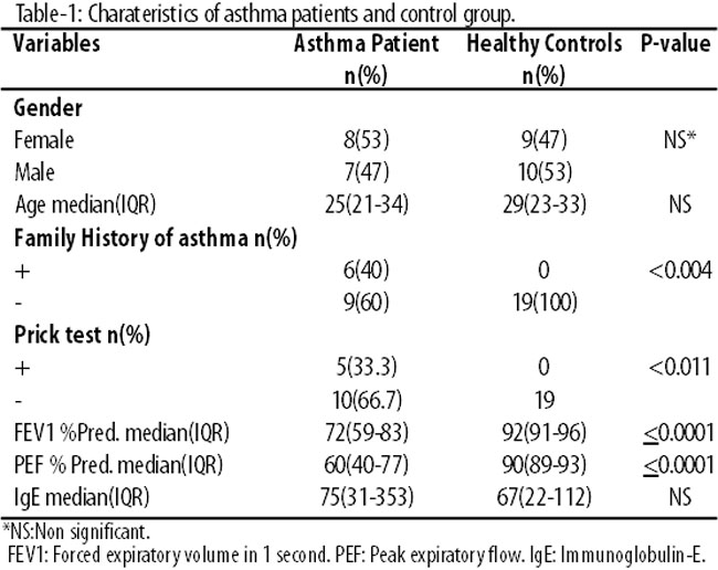
Pre- and post-treatment FEV1 and PEF values were compared and found to be increased after the therapy (Table-2).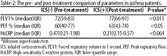
However, there was no statistically significant change in mean serum hsCRP levels before or after therapy (p<0.064).
The mean hsCRP level in the Control group was 0.29±0.24mg/dL (median: 0.26; 25-75% quartiles 0.086-0.434). The level of hsCRP in the Case group prior to treatment was significantly higher than the Control group (p=0.036). Statistical difference was not observed between the post-therapy and the Control group hsCRP levels (p=0.357) (Figure-1).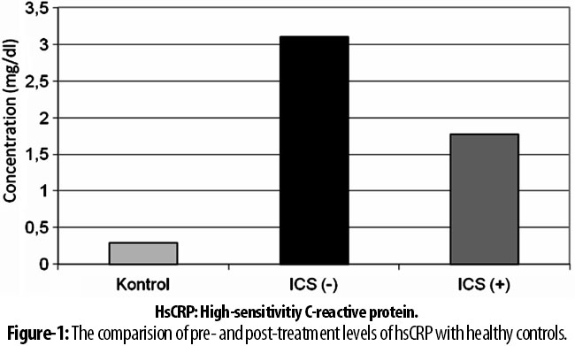
No significant correlation was seen between the pre-treatment PFT measurements and serum hsCRP levels (r=-0.289, p<0.390 and r=0.03, p<0,917) (Table-3).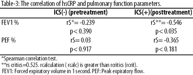
However, the post-treatment FEV1 measurement was negatively correlated with hsCRP levels (r=-0.546, p< 0.035) (Figure-2).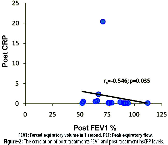
The correlation between positive prick tests and total IgE levels was not determined.
Discussion
Several studies have demonstrated that there is systemic inflammation in addition to local inflammation in asthmatic patients. CRP is the defining marker of inflammatory processes. Recently, it has been reported that hsCRP, which traditionally is used as an indicator of low level systemic inflammation, is also a good maker for inflammation in asthma.1,8
However, some studies did not show statistical differences in CRP levels between asthma and healthy control groups.2
This study was performed with patients who were newly diagnosed with moderate asthma, who were not on asthma medications. HsCRP was found to be useful in detecting low levels of inflammation due to excellent sensitivity and favourable kinetics. The pre- and post-corticosteroid hsCRP levels were determined in order to study the early effect of inhaled corticosteroid therapy on hsCRP levels.
Serum hsCRP levels were significantly higher in asthmatics prior to the treatment compared to controls, suggesting that systemic inflammation is present in asthma. The average hsCRP level declined following the therapy, but the difference was not statistically significant. Additionally, post-therapy hsCRP levels were not significantly different than controls.
In an important study, it was observed that increasing levels of hsCRP were associated with decreased PFT measurements in untreated mild asthma patients compared to the control group.2 It concurrently demonstrated that there was no difference in the level of serum and exhaled hsCRP between patients treated with and without steroid. However, the steroid and non-steroid groups were composed of separate patients, rather than comparing values from the same patients on and off therapy. Serum and exhaled hsCRP levels were increased in asthmatics compared to the controls.8 Though the methodology was different in comparing pre- and post-treatment, our findings are consistent with literature.
In one study, patients with asthma and no history of steroid therapy had increased serum hsCRP levels compared to the controls. It was emphasised that hsCRP might serve as surrogate marker of airway inflammation in asthma.15 HsCRP were gradually increased in children with asthma from mild degree to moderate and severe degree in another study.17 In our study, no difference in serum hsCRP levels was seen after one month of corticosteroid therapy compared to the controls. However, serum hsCRP levels were statistically significant in asthma patients prior to the therapy compared to the controls. The data suggests that corticosteroid therapy may supress inflammation. Our study was limited by the small number of patients. This study was designed as a pilot study and the results suggest that we should continue the study in the future.
One study did not determine any statistical difference between patients treated with corticosteroids and healthy controls. However, in this study, which included 22 patients who were treated with inhaled corticosteroids, the asthma severity of patients was not specified.15 Another demonstrated a reduced level of hsCRP with corticosteroid therapy in a greater number of patients, but did not clarify the level of severity of the asthma patients. It evaluated the effects of prolonged therapy (duration greater than 3 months).14 Some studies have shown correlations between increased hsCRP levels, decreased FEV1 values and increased airway hyper-responsiveness.10,18 We did not find a correlation between hsCRP and PFT measurements prior to the therapy because all the patients had airway obstruction prior to steroid treatment. Following corticosteroid treatment, a negative correlation between FEV1 and hsCRP levels was seen. This result may be because corticosteroid therapy decreased hsCRP levels and improved pulmonary functions parametres. This correlation may be due to different baseline hsCRP values and individual responsiveness to corticosteroid therapy, but this conclusion needs to be verified by further studies. In contast to one study,10 another reported that hsCRP was not a good marker of bronchial hyper-reactivity.19 In the latter study, suspected asthma patients with either positive or negative bronchial hyperreactivity to histamine challenge were evaluated. High levels of hsCRP were observed in patients with positive reactions to histamine. This finding could have been due to the fact that short-term, local inflammation was not reflected in systemic circulation. In this study, only moderate asthmatic patients were included. Therefore, it is expected that markers of inflammation would be present even at low levels of inflammation. A decrease in serum hsCRP was expected after the therapy. However, this decrease was not observed, possibly because of the short duration of therapy or because of the small number of patients. HsCRP may have more utility in determining the response to treatment for severe asthma patients.20 In a study, serum hsCRP levels were significantly higher in uncontrolled steroid-naïve asthmatics than those controlled on inhaled steroid with a median of 3.15µmg/L and 1.55µmg/L, respectively. They observed serum hsCRP had sensitivity of 72% and a specificity of 93%.21
Patients with conditions other than asthma that are known to influence the level of hsCRP were excluded from our study. For instance, it is well known that CVD, DM, obesity and smoking lead to increased CRP levels.22,23 Therefore, all of our patients were examined by an Internal Medicine specialist and cardiologist to rule out these diseases.
Additionally, aging also can contribute to high hsCRP.22 Only young, non-obese, non-smoker asthmatics with and no other underlying causes of inflammation were included in the study.
HsCRP levels are strongly predictive of cardiovascular events, including myocardial infarction, ischaemic stroke and peripheral vascular diseases.23,24 The utility of this biomarker for cardiovascular risk stratification in patients with and without established CVD is supported by evidence.25 In this study, high levels of hsCRP were found in young patients with moderate, rather than severe, asthma. Prolonged elevated hsCRP levels may increase the risk of CVD. Furthermore, inflammatory markers in asthma can circulate throughout the body and cause vascular injury.
The exact function of CRP in humans is unclear. This protein has been shown to participate in inflammatory as well as innate immunity processes. The bioactivity of CRP is determined by its ability to bind a variety of ligands, such as damaged cell membranes, apoptotic cells, fibronectin, etc, with the highest affinity for phosphocholine residues. CRP is ligand bound and activates the classical complement pathway. CRP is also a regulator of the alternative complement pathway. CRP was originally categorised as a liver-derived protein, but recent evidence shows significant levels of CRP in blood vessels and coronary artery smooth muscle cells. Taken together, these findings suggest that CRP is not only a marker, but also a potential contributor to inflammatory diseases.6,26-28 Novel understanding of the localised dissociation of circulating pentameric CRP to monomeric forms demonstrates new aspects of the inflammatory process and further highlights this process as a potential therapeutic target.28,29 Therefore, long-term follow-up of patients diagnosed at an early stage should be done. Anti-inflammatory therapy should be started as early as possible to decrease cardiovascular risk. However, studies that are designed to compare patients in the early, middle and late periods of corticosteroid therapy who have different severity levels of asthma are needed. Recently, a study reported that levels of serum hsCRP could identify distinct aspects of local and systemic inflammation in patients with obstructive airways disease. However, hsCRP may elevate in COPD patients, too. Routine measurements of hsCRP, IgE, and blood eosinophils are proposed for the diagnosis of asthma and COPD in a primary care setting to provide information about the response to treatment or disease progression.30
Conclusion
HsCRP is a marker of systemic inflammation in moderate asthma. Future studies with a more diverse level of asthma control are warranted in examining the utility of hsCRP in the evaluation of asthma. Besides, long-term studies with larger number of patients are also needed to evaluate the relationship between hsCRP and cardiovascular indications.
References
1. Busse WW, Lemanske RF Jr. Asthma. N Engl J Med 2001; 344: 350-62.
2. Büyüköztürk S, Gelincik AA, Genç S, Koçak H, Öneriyidogan Y, Erden S, et al. Acute phase reactions in allergic airway disease. Tohoku J Exp Med 2004 ; 204: 209-13.
3. Jousilahti P, Salomaa V, Hakala K, Rasi V, Vahtera E, Palosuo T. The association of sensitive systemic inflammation markers with bronchial asthma. Ann Allergy Asthma Immunol 2002; 89: 381-5.
4. Tall AR. C-reactive protein reassessed. N Engl J Med 2004; 350:1450-2.
5. Pradhan AD, Manson JE, Rifai N, Buring JE, Ridker PM. C-reactive protein, interleukin 6, and risk of developing type 2 diabetes mellitus. JAMA 2001; 286: 327-34.
6. Bard RL, Clarke N, Rubenfire M, Eagle K, Brook RD. P-594: cardiovascular risk stratification obtained by Framingham risk score and C-reactive protein measurement. Am J Hypertens 2005; 18: 224A
7. Rifai N, Tracy RP, Ridker PM. Clinical efficacy of an automated high-sensitivity C-reactive protein assay. Clin Chem 1999; 45: 2136-41.
8. Zietkowski Z, Tomasiak-Lozowska MM , Skiepko R, Mroczko B, Szmitkowski M, Bodzenta-Lukaszyk A. High-sensitivity C-reactive protein in the exhaled breath condensate and serum in stable and unstable asthma. Respir Med 2009;103: 379-385.
9. Fujita M, Ueki S, Ito W, Chiba T, Takeda M, Saito N, et al. C-reactive protein levels in the serum of asthmatic patients. Ann Allergy Asthma Immunol 2007; 99: 48-53.
10. Kony S, Zureik M, Driss F, Neukirch C, Leynaert B, Neukirch F. Association of bronchial hyperresponsiveness and lung function with C-reactive protein (CRP): a population based study. Thorax 2004; 59: 892-6.
11. Olafsdottir IS, Gislason T, Thjodleifsson B, Olafsson I, Gislason D, Jögi R, et al. C- reactive protein levels are increased in non-allergic but not allergic asthma: a multicentre epidemiological study. Thorax 2005; 60: 451-54.
12. Azar RR, Rinfret S, Théroux P, Stone PH, Dakshinamurthy R, Feng YJ, et al. A randomized placebo-controlled trial to assess the efficacy of antiinflammatory therapy with methylprednisolone in unstable angina (MUNA trial). Eur Heart J 2000; 21: 2026-2032
13. Versaci F, Gaspardone A, Tomai F, Ribichini F, Russo P, Proietti I, et al. Immunosuppressive therapy for prevention of restenosis after coronary artery stent implantation (IMPRESS Study). J Am Coll Cardiol 2002; 40:1935-1942
14. Kasayama S, Tanemura M, Koga M, Fujita K, Yamamoto H, Miyatake A. Asthma is an independent risk for elevation of plasma C-reactive protein levels. Clin Chim Acta 2009; 399: 79-82
15. Takemura M, Matsumoto H, Niimi A, Ueda T, Matsuoka H, Yamaguchi M, et al. High sensitivity C-reactive protein in asthma. Eur Respir J 2006; 27: 908-12.
16. GINA Report, Global Strategy for Asthma Management and Prevention 2010(Updated). (Online) (Cited 2011 February 5). Available from URL: http://www.ginasthma.org.
17. Wu SH, Yin PL, Zhang YM, Tao HX. Reversed changes of lipoxin A4 and leukotrienes in children with asthma in different severity degree. Pediatr Pulmonol 2010, 45: 333-340.
18. Fogarty AW, Jones S, Britton JR, Lewis SA, McKeever TM. Systemic inflammation and decline in lung function in a general population: a prospective study. Thorax 2007;62:515-20.
19. Panaszek B, Liebhart E, Liebhart J, Pawlowicz R, Fal AM. Serum concentration of C-reactive protein is not a good marker of bronchial hyperresponsiveness. Arch Immunol Ther Exp (Warsz) 2007; 55: 341-5.
20. Qian FH, Zhang Q, Zhou LF, Liu H, Huang M, Zhang XL, et al. High-sensitivity C-reactive protein: a predicative marker in severe asthma. Respirology 2008; 13: 664-669.
21. Deraz TE, Kamel TB, El-Kerdany TA, El-Ghazoly HM, High-sensitivity C reactive protein as a biomarker for grading of childhood asthma in relation to clinical classification, induced sputum cellularity, and spirometry. Pediatr Pulmonol 2012, 47: 220-225.
22. Ridker PM. High-sensitivity C-reactive protein: potential adjunct for global risk assessment in the primary prevention of cardiovascular disease. Circulation 2001 3;103:1813-8.
23. Yudkin JS, Stehouwer CD, Emeis JJ, Coppack SW. C-reactive protein in healthy subjects: associations with obesity, insulin resistance, and endothelial dysfunction: a potential role for cytokines originating from adipose tissue? Arterioscler Thromb Vasc Biol 1999;19:972-8.
24. Nakamura H, Yamashita T. Predictive value of cardiovascular events by high sensitivity CRP. Nihon Rinsho 2002; 60: 916-21.
25. Sabatine MS, Morrow DA, Jablonski KA, Rice MM, Warnica JW, Domanski MJ, et al. Prognostic significance of the Centers for Disease Control / American Heart Association high-sensitivity C-reactive protein cut points for cardiovascular and other outcomes in patients with stable coronary artery disease. Circulation 2007; 115: 1528-36.
26. Calabrò P, Golia E, Yeh ET. CRP and the risk of atherosclerotic events. Semin Immunopathol 2009; 31: 79-94.
27. Diehl EE, Haines GK 3rd, Radosevich JA, Potempa LA. Immunhistochemical localization of modified C-reactive protein antigen in normal vascular tissue. Am J Med Sci 2000; 319:79-83
28. Bíró A, Rovó Z, Papp D, Cervenak L, Varga L, Füst G, et al. Studies on the interactions between C-reactive protein and complement proteins. Immunology 2007; 121:40-50.
29. Eisenhardt SU, Thiele JR, Bannasch H, Stark GB, Peter K. C-reactive protein: how conformational changes influence inflammatory properties. Cell Cycle 2009; 8: 3885-92
30. Tilemann L, Gindner L, Meyer F, Szecsenyi J, Schneider A. Differences in local and systemic inflammatory markers in patients with obstructive airways disease. Prim Care Respir J 2011; 20: 407-14.
Journal of the Pakistan Medical Association has agreed to receive and publish manuscripts in accordance with the principles of the following committees:


