

Saki Ghasem ( Phsiology Research Center, Ahvaz Jundishapur University of Medical Sciences, Ahvaz. Iran. )
Jasemi Majid ( Department of Urology, Golestan Hospital, Faculty of Medicine, Ahvaz Jundishapur University of Medical Sciences, Ahvaz. Iran. )
Razi Shiva ( Phsiology Research Center, Ahvaz Jundishapur University of Medical Sciences, Ahvaz. Iran. )
July 2013, Volume 63, Issue 7
Original Article

Abstract
Objective: To assess developmental capacity of fertilised oocytes by sperm of mouse exposed to forced swimming stress.
Methods: The experimental study was conducted at the Physiology Research Center of Ahvaz Jundishapur University of Medical Sciences, from August 2011 to January 2012. It comprised 20 adult male and 10 female mice. The male mice were randomly divided into two equal groups (n=10): control and experimental. Animals of the experimental group were submitted to forced swimming stress. All male mice were euthanised and the cauda epididymis removed before contents were squeezed out. A pre-incubated capacitated sperm was gently added to the freshly collected ova of the two groups of study. The combined sperm-oocyte suspension was incubated for 4-6 hours under a condition of 5% Carbon dioxide and 37°C temperature. The ova were then washed through several changes of medium and finally incubated. Fertilisation was assessed by recording the number of 1-cell embryos 4-6 hours after insemination. The 1-cell embryos were allowed to further develop in vitro for about 120 hours. Development of embryos everyday and during 5 days of culture was observed by using inverted microscope. SPSS 13.0.1 was used for statistical analysis.
Results: The percentage of oocytes fertilised was 75:96 (78.12±4.8%) and 50:10 (49.5±3.9%) in the control and experimental groups, respectively. The difference was significant (p <0.001). At 24 hours after insemination, 70:75 (93.33±2.7%) and 39:50 (78±3.5% )of fertilized oocytes developed to two=cell embryos in control and experimental groups respectively. The difference was significant (p <0.02). There were not significant differences (p>0.05) between the two groups in terms of speed and developmental capacity of blastocysts.
Conclusions: Fertilisation capacity of male mice affected by forced swimming stress and also the developmental capacity of oocyte fertilised by sperm of mouse exposed to forced swimming stress decreased.
Keywords: Oocyte, Infertility, Stress, Mouse. (JPMA 63: 838; 2013).
Introduction
Infertility is defined as the inability of a couple to conceive a pregnancy after attempts across one full year. Physical and psychological factors can cause infertility. Infertile couples may suffer from female factors, male factors, or both.1 Psychological stress is commonly recognized as a contributing factor to infertility and has been found to be high in infertile couples. Psycho-amenorrhea is more seen in women with stressful lives and situations, are usually underweight, single and they used to use psycho-active drugs.2-4 Many studies have investigated psychological causes that affect male factor infertility,5-9 like sperm count, motility and morphology. Previous studies shown impotence, sham ejaculation and oligospermia to be associated with psychological factors in male infertility.10,11 A wide range of stress factors such as micro-organisms, hyperthermia and exposure to heavy metals affect reproductive functions and sperm production in males.12 Previously, many researchers have declared that same effects were observed after implementation of the stressful stimuli such as prolonged immobilisation and forced swimming stress.13,14 In a previous study, it was demonstrated that forced swimming is one of the stressful factors in the time course just like spermatogenesis period.15 Previous study showed that the rate of pre- and post-implantation loss in female rats mated with stressed male rats obviously increased.16,17 After literature review, we found no published report on the effect of forced swimming stress on the developmental capacity of mouse oocyte fertilised by sperm of mouse exposed to forced swimming stress. The present study was designed to address the subject.
Material and Methods
The experimental study was conducted at the Physiology Research Center of Ahvaz Jundishapur University of Medical Sciences, Ahvaz, Iran, from August 2011 to January 2012. A total 20 of adult Noval Medical Research Institute (NMRI) male mice 10-12 weeks of age with a mean weight of 50±3.6g were purchased from the Laboratory Animals Care and Breeding Center of the university. The mice were randomly divided into two equal groups of 10 Control and 10 Experimental mice. They were individually housed in cages under a 12-h light/dark cycle, at 23±3°C temperature and 60-65% humidity-controlled room with free access to food and water. All procedures were approved by international guidelines and by the Institute Research Ethics and Animal Care and Use Committee of the university. Every effort was made to minimize the number of animals used and their suffering.
The control group remained in the cages, while the experimental mice were submitted to forced swimming for 3 minutes in water at 32°C daily for 50 days. Stress was assessed by the hot-plate test after the last day.14 In the hot-plate, the maximal cutoff time was 60 seconds and temperature was 52°C. The latency time for the hind licking after exposure to the hot-plate surface was measured and the increase in relation to Controls was considered to be an index of the anti-nociceptive effect.
For oocyte collection 10 NMRI females were administered intra-peritoneally 5 IU pregnant mare serum gonadotropin (PMG) for super-ovulation. This was followed 46-48 hours later by the intra-peritoneal administration of 5 IU human chorionic gonadotropin (HCG). About 12-14 hours after the HCG injection, the mice were killed by cervical dislocation method and the oviducts were excised before cumulus-oocyte complexes were collected in K± modified simplex optimized medium (KSOM) medium. The granulosar cells of oocytes were removed by repeated pipetting in KSOM medium containing 80 IU.ml-1 hyaluronidase and the mature oocytes thus obtained were divided in two groups randomly.
Males in the two groups were sacrificed before right cauda epididymis was removed, and immediately inserted into a drop of Toyoda, Tokoyama, Mosi (TYH) medium+5 mg. mL-1 Bovine Serum Albumin (BSA) under mineral oil (Sig., embryo-tested, cat. No. M8410). Spermatozoa were extracted from the medium and incubated for 1 hour 37°C in 5% carbon dioxide (CO2) in humidified air for the purpose of capacitation.18,19
In vitro fertilisation was carried out in drops of KSOM medium plus 5mg/ml-1 Bovine Serum Albumin (BSA) under mineral oil. A pre-incubated capacitated sperm was gently added to the freshly-collected ova, then incubated for 4-6 hours under conditions of 5% CO2 and 37°C temperature. Ova was then washed through several changes of the medium. Fertilization was assessed by recording the number of 1-cell embryos (2PN) 4-6h after insemination.20
The 1-cell embryos were allowed to further develop in vitro for about 120 hours in the same medium. Development of embryos every day and during 5 days of culture was observed by using inverted microscope. Embryos with 25% or more fragmentation with opaque cytoplasm were considered degenerated and were excluded from the study.
For data purposes, each drop was considered as an experimental unit. The fertilisation rate of oocyte was obtained by counting of 1-cell embryo in all drops of every group of study. Mean ± standard deviation of cleaved embryos or embryo that developed to another step was obtained after calculating the percentage of cleaved embryo in every drop of each group. The statistical significance of difference between the control and experimental groups was determined by the student\\\'s t-test. Statistical analysis was performed using SPSS version 13.0.1. Differences were considered significant when p<0.05 was achieved.
Results
The 196 extracted oocytes were divided into two groups: 96 (48.73%) as control, 101 (51.26%) as experimental. The percentage of oocytes that fertilised was 75 (78%) and 50 (49%) in the two groups respectively (p <0.001). The percentage of embryo in morula stage was significantly increased (p <0.05) in the control group, while the rate of 4-cells embryos in the experimental group was significantly high (p <0.003) in compared to control group (Table-1).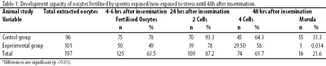
The development of 4-cell embryos to the blastocyst stage was also evaluated (Table-2).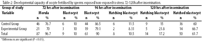
The 4 cell embryos of the two groups could reach to blastocyst stage at 72 hours after insemination. There was no difference in percentage of embryos in the blastocyst stage of the two groups; of 6 (10%) in Control and 3 in Experimental group (p>0.05). On examining the developmental capacity and the speed of development of blastocysts at 96 and 120 hours after insemination, it was observed that the blastocysts developed hatching balstocyst and hatched stage (advanced stage of development). There were no significant differences (p>0.05) between the two groups in terms of speed and developmental capacity.
Discussion
Previously studies have shown the decrease in male fertility during the last few decades due to harmful environmental influences, pollutants and stress.4 The current study also showed that, the stress caused by forced swimming decreased in vitro fertilisation capacity. The decreased fertilisation capacity of the stressed male may be due to decrease in sperm motility. In an earlier study, it was demonstrated that forced swimming stress affected the motility of sperms.15 The current study observed that the fertilised oocytes of the two groups could develop to two-cells, four-cell embryos and morula at 24, 48 and 72 hours after insemination, but the rate of cleavage and speed of development in the control group was significantly higher than the experimental group. It may be because fertilisation of oocytes with injured spermatozoa. There are some reports about the deleterious effect of stress on animal and human semen quality, like decrease in seminal volume, sperm concentration and motility, and in the number of normal spermatozoa.19,21 A study observed that the sperm dioxyribonucleir acid (DNA) contribute one half of the genomic material of the offspring. As such, normal sperm genetic material is required for fertilisation, embryo and foetal development and post-natal child well-being.22 To our knowledge the effect of forced swimming stress on DNA has not been studied and reported yet. It is now up to those topic of stress and its effects on the genetic structure of sperm to conduct further investigations. The current study examined the speed and developmental capacity of blastocysts at 4 to 5 days after insemination and observed no significant difference between the two groups. Indeed, the reason for our findings is not clear, but probably it is related to degenerated and bad quality embryos in the early stage, which means that in the final stages only healthy embryos survive. There may be other reasons which need to be looked into.
Conclusion
The fertilisation capacity of male mice was affected by forced swimming stress, and the speed and development capacity of oocyte fertilised by sperm of mouse exposed to forced swimming stress also decreased.
Acknowledgements
We are grateful to the Research Deputy of Ahvaz Jundishapur University of Medical Sciences for the financial support, and to Miss Iqbal Zakerhosseini for editorial assistance.
References
1. Bhattacharya S, Porter M, Amalraj E, Templeton A, Hamilton M, Lee AJ, et al. The epidemiology of infertility in the North East of Scotland. Hum Reprod 2009; 24: 3096-107.
2. Shahid S. Depression in infertile couples. J Coll Physicians Surg Pak 2009; 19: 395-6.
3. Wischmann T, Scherg H, Strowitzki T, Verres R. Psychosocial characteristics of women and men attending infertility counselling. Hum Reprod 2009; 24: 378-85.
4. Kaneko M, Ali M, Ushijima H. Relationship between decreasing fertility during the post-war period and maternal age in a Japanese population. Tohoku J Exp Med 2004; 202: 221-6.
5. Zorn B, Auger J, Velikonja V, Kolbezen M, Meden-Vrtovec H. Psychological factors in male partners of infertile couples: relationship with semen quality and early miscarriage. Int J Androl 2008; 31: 557-64.
6. Clarke RN, Klock SC, Geoghegan A, Travassos DE. Relationship between psychological stress and semen quality among in-vitro fertilization patients. Hum Reprod 1999; 14: 753-8.
7. Hjollund NH, Bonde JP, Henriksen TB, Giwercman A, Olsen J. Job strain and male fertility. Epidemiology 2004; 15: 114-7.
8. Sheiner E, Sheiner EK, Potashnik G, Carel R, Shoham-Vardi I. The relationship between occupational psychological stress and female fertility. Occup Med (Lond) 2003; 53: 265-9.
9. Pook M, Tuschen-Caffier B, Kubek J, Schill WB, Krause W. Personality, coping and sperm count. Andrologia 2005; 37: 29-35.
10. Palti Z. Psychogenic male infertility. Psychosom Med 1969; 31: 326-30.
11. Apfel RJ, Keylor RG. Psychoanalysis and infertility. Myths and realities. Int J Psychoanal 2002; 83: 85-104.
12. Ozawa N, Goda N, Makino N, Yamaguchi T, Yoshimura Y, Suematsu M. Leydig cell-derived heme oxygenase-1 regulates apoptosis of premeiotic germ cells in response to stress. J Clin Invest 2002; 109: 457-67.
13. Almeida SA, Kempinas WG, Lamano Carvalho TL. Sexual behavior and fertility of male rats submitted to prolonged immobilization-induced stress. Braz J Med Biol Res 2000; 33: 1105-9.
14. Mingoti GZ, Pereira RN, Monteiro CM. Fertility of male adult rats submitted to forced swimming stress. Braz J Med Biol Res 2003; 36: 677- 81.
15. Saki G, Rahim F, Alizadeh K. Effect of forced swimming stress on count, motility and fertilization capacity of the sperm in adult rats. J Hum Reprod Sci 2009; 2: 72-5.
16. Saki G, Rahim F, Vaysi OA. Effect of forced swimming stress on in-vivo fertilization capacity of rat and subsequent offspring quality. J Hum Reprod Sci 2010; 3: 32-4.
17. Saki G, Razie S, Amirpoor S. Pregnancy rate in female mice exposed to forced swimming stress. Asian J Biol Sci 2011; 4: 266-71.
18. Giblin PT, Poland ML, Moghissi KS, Ager JW, Olson JM. Effect of stress and characteristic adaptability on semen quality in healthy men. Fertil Steril 1988; 49: 127-32.
19. Almeida AS, Petenusci SO, Anseimo-Franci JA, Rosa-e-Silva AA, Lamano-Carvalho TL. Decreased spermatogenic and androgenic testicular functions in adult rats submitted to immobilization-induced stress from prepuberty. Braz J Med Biol Res 1998; 31: 1443-8.
20. Jalali M, Saki G, Sarkaki AR, Karami K, Nasri S. Effect of noise stress on count, progressive and non-progressive sperm motility, body and genital organ weights of adult male rats. J Hum Reprod Sci 2012; 5: 48-51.
21. Steinberger E. The etiology and pathophysiology of testicular dysfunction in man. Fertil Steril 1978; 29: 481-91.
22. Nili HA, Mozdarani H, Aleyasin A. Correlation of sperm DNA damage with protamine deficiency in Iranian sub-fertile men. Reprod Biomed Online 2009; 18: 479-85.
Journal of the Pakistan Medical Association has agreed to receive and publish manuscripts in accordance with the principles of the following committees:


