

Yousuf A. Husen ( Department of Radiology, Aga Khan University Hospital, Stadium Road, Karachi. )
Tahir R.Khalid ( Department of Radiology, Aga Khan University Hospital, Stadium Road, Karachi. )
Zahid A. Khan ( Department of Radiology, Aga Khan University Hospital, Stadium Road, Karachi. )
M.Y Sheikh ( Department of Radiology, Aga Khan University Hospital, Stadium Road, Karachi. )
November 1997, Volume 47, Issue 11
Original Article

Abstract
To assess the lack of visibility of vascular markings under the hemidiaphragm on a frontal chest radiograph as a sign of pleural effusion, fifteen patients were collected showing this sign. Pleural effusion was diagnosed by ultrasound, comparison with previous or subsequent chest x-ray or computed tomography. Patients in the study group exhibited this sign in the absence of the classical signs of pleural effusion. In the control group, lack of visibility of blood vessels was observed in only 4.2% cases. Non-visualization of vascular markings below the hemidiaphragm should alert the interpreter to the possible presence of pleura! effusion and a lateral or decubitus view or ultrasound examination may be carried out to rule out effusion (JPMA 47:284,1997).
Introduction
Pleural effusion is one of the most common positive findings noticed on a chest radiograph and its value thus cannotbe overemphasized. Early detection of pleural effusion has been described by many authors1-4. Schwarz described a new radiologic sign of subpulmonic effusion as obliteration of normally seen intrapulmonaiy blood vessels below thelevel of hemidiaphragm3. In routine practice we have observed many such cases in which the only clue to the presence of subpulmonic effusion on frontal chest x-ray was lack of visibility of lung markings below hemidiaphragm. This report is based on 15 such cases.
Patients and Methods
The study gmup comprised of 17 subpulmonic effusions in 15 patients without typical chest roentgenogmphic signs to prompt the diagnosis, Two patients had bilateral involvement. Non-visualization of lung markings below the hemidiaphragm was the basis of early detection of subpulmomc effusion. All films were taken at high kVp (100-110) and low mAs (2-5) technique. The fmntal erect or (supine) chest radiogmphs of these patients were evaluated for the presence of occult signs of pleural effusions i.e., loss of lung markings below the hemidiaphragm and increased density below the hemidiaphragm on the affected side. Interpretation was done by two radiologists (YAH and TRK) independently. The diagnosis of pleural effusion was confinned by ultrasound (US), computed tomography (CT), decubitus view or comparison with previous or subsequent chest film demonstrating normal vascular visibihty below the hemidiaphragm in question. Underexposed films, patients with significantobesity or large breasts obscuring the region of interest and those with known ascites were excluded from the study. Visibility of vessels below hemidiaphragm was assessed in 105 normal subjects as control gmup.
Results
The age range of the study group subjects was 9 to 68 years with a male to female ratio of 3:2, The control gmup had an age range of 16 to 80 years and a male to female ratio of 1:1.9. Of the 17 subpulmonic effusions, lateral costophrenic sulcus was sharp and pointed in configuration in 14, while changed equivocally (minimally blunted, hazy or displaced) in remaining 3.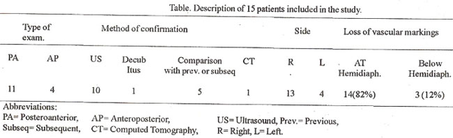
Table gives description of patients used in the study alongwith,mode of confirmation of pleural effusion and pattern of loss of vascular markings below the hemidiaphragm. In 5 patients comparison with previous or follow-up films, revealing normal subdiaphiagmatic vascular pattern, was used as a diagnostic sign for confirmation of pleural effusion (figure la and ib).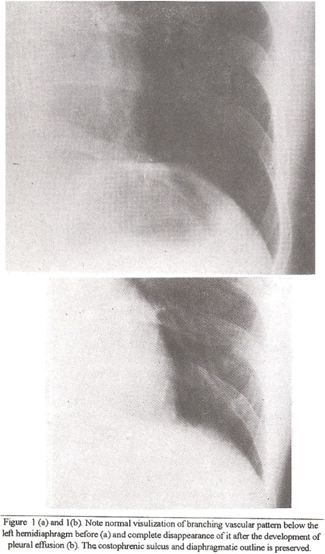
Vascularvisibility endedat (n=14) or just below (n=3) the level of presumed hemidiaphragm. Three patients in latter group showed abrupt change in the vascular visibility witha sharp cutoff. Incontrol group, the distance for which vessels were seen coursing below the diaphragm rangedfromO to 8.5 cmon right and 0 to 5.5 cm on left. Mean length of visualized vessels was 4.3 cm on right and 2.4cm on left. Typically a gradual loss ofvascular visibility was noted from above downwards. In 9 normal cases (4.2% hemidiaphragms) vessels were not visualizedbelow the hemidiaphmgm(8 on left and 1 onnght).
Discussion
Blunting of lateral costophrenic sulcus, loss of hemidiaphragm, increased density below hemidiaphragm and meniscus signs are classical manifestations of pleural effusion. Subpulmonic effusions may be diagnosed by raised hemidiaphragm, straightening of hemidiaphragm and Hessel’s sign i.e., lateral shift of the superior most portion of hemidiaphragm on the frontal chest film2. Detection of minute subpulmonic effusion may be difficult on the frontal film. Whilst the decubitus examination can detect as little as 5-15 ml of pleural fluid1, it may take 200-600 ml of fluid to cause blunting of the lateral costophremc sulcus2,5 to be seen on frontal examination.Minute fluid collection starts in the subdiaphragmatic location, spilling to posterior costophrenic sulcus, obliteration of which may be seen only on the lateral view8. Frontal film, which could be the only examination available inroutine cases, may notshow aconclusive evidence of subpulmonic effusion in these cases. Schwarz described 3 cases with loss of vascular visibility below hemidiaphragm as the only sign of pleural effusion3. Present study included 12 such patients. The remaining 3 had minimal equivocal vascular change observed included loss of visibility at (82%) orbelow (18%) the presumed hemidiaphragm.
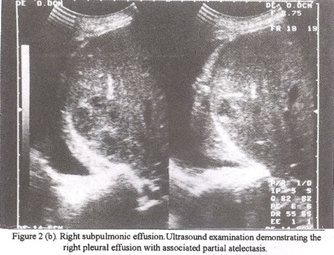
Comparison of previous or subsequent films is useful in the early detection of this vascular divergence. If on one occasion the vessels below hemidiaphragm are discernible anda comparable frontal film on anotheroccasion reveals loss of visibility, the possibility of subpulmonic effusion should be strongly suggested. This criteria was used successfully in 5 patients (Figure laand ib). The signwas seen more frequently on right side probably because of more homogenous backgroundprovidedby liver leading to better appreciation of vascular pattern as compared to left side where overlapping bowel shadows interfere with visibility of already hard to see “subdiaphragmatic” vessels. Other conditions which may obscure vessels below hemidiaphragm due to increased density or basal lung disease, include basal consolidation or collapse, subdiaphragmatic pathology, large breasts and obesity3,6. The fmal diagnosis should therefore be entertained in the light of clinical picture and associated radiologic signs. It is postulated that the blood vessels am obscured by two situations: a) increased density produced by the pleural effusion; b) compression of adjacent lung that commonly accompanies pleural effusion6 (Figure 2a and 2b).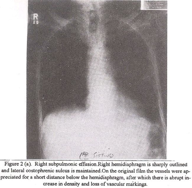
Loss of or abrupt change in vascular visibility below diaphragm may be the only sign in minute subpulmonic effusion. Wheneverseen, itshouldpromptfurtherevaluationby ultrasound examination or decubitus film. Comparison with previous or subsequent films showing normal vascularity in the region may also be of help.
References
1. Harold, M., Robert, TP., Ronald, S. et al. Roentgen visualization of minute pleural effusion. Radiology, 1973; 109:33- 36.
2. Rudikoff.J.C. Early detection ofpleural fluid. Chest, 1980;77:109-11 1.
3. Marwin, IS. and Barry. L.M. A new radiologic sign ofsub- pulmonic effusion. Chest, 1975;67:1 76-178.
4. Rigler, L.G. Roentgen diagnosis of small pleural effusion. A new roentgenographic position. JAMA, 1931 ;96: 104.
5. Bowen, A. Quantitative roentgen diagnosis of pleural effusions. Radiology, 1931;17:520.
6. Fraser, R.G. and Pare, JAR Diagnosis of diseases of the chest. Vol 1. Philadelphia, London, Toronto, WB. Saunders, 1977, p. 575.
Journal of the Pakistan Medical Association has agreed to receive and publish manuscripts in accordance with the principles of the following committees:


