

Muhammad Azeemuddin ( Department of Radiology, Aga Khan University Hospital, Karachi. )
Rohana Naqi ( Dow University of Health Sciences, Karachi. )
Mohammad Wasay ( Aga Khan University Hospital, Karachi. )
June 2013, Volume 63, Issue 6
Original Article

Abstract
Objective: To explore equation, if any, between findings of magnetic resonance imaging of pituitary gland and serum prolactin level.
Methods: The retrospective, descriptive study was conducted at the Department of Radiology, Aga Khan University Hospital, Karachi, and related to patients\\\' records from April 19, 2006 to April 23, 2009. Seventy patients underwent magnetic resonance imaging of brain for pituitary gland. Inclusion criteria were all patients referred with relevant clinical symptoms or deranged serum prolactin level. Patients who were claustrophobic or had a pacemaker, aneurysm clip, metallic foreign body in the orbit or with no laboratory investigation were excluded from the study. SPSS 19 was used for statistical analysis.
Results: Of the 70 patients, normal imaging was noted in 29 (41.4 %) patients. Out of these, 18 (62.06%) patients had normal and 11 (37.93%) had raised serum prolactin levels. Microadenoma was found in 23 (32.8%) patients. Out of these, 10 (42.47%) had normal and 13 (56.52%) had raised prolactin levels. Macroadenoma was found in 16 (22.8 %). Out of these, 8 (50%) had normal and 8 (50%) had raised prolactin levels. Pituitary cyst was located in 2 (2.8 %) patients. Out of these, 1 (50%) had normal and 1 (50%) had raised serum prolactin levels.
Conclusion: Magnetic resonance imaging of pituitary gland was not associated with serum prolactin levels in patients with clinical suspicion of pituitary abnormality. Therefore, regular monitoring of serum prolactin is suggested.
Keywords: Magnetic resonance imaging, Pituitary adenoma, Serum prolactin level. (JPMA 63: 739; 2013).
Introduction
Magnetic resonance imaging (MRI) is now used routinely in the investigation of the pituitary gland, and is accepted as the most sensitive imaging method for the diagnosis of pituitary microadenomas.1 In symptomatic patients with galactorrhoea, menstrual irregularities, amenorrhoea, or impaired reproductive function, prolactin disorders should be suspected and pituitary adenoma should be sought. Reported sensitivities of MRI in the detection of microadenomas vary widely, but are about 85-90% for contrast-enhanced studies.2 The diagnosis of prolactin-secreting pituitary adenoma is made on clinical and endocrinological evidence. The role of radiology is to assess the size of the adenoma (to assess the effects of therapy), and possibly the feasibility of surgery. Though computed tomography (CT) was thought to be extremely useful in the diagnosis of pituitary microadenomas,3 high-resolution MRI has become the most sensitive means for their detection.4 Previous studies suggest a high rate of detection.5
A study of 74 patients in 1996, with serum prolactin levels above 52ng/ml found microadenomas in 38 (51.3%) patients, macroadenomas in 6 (8.1%) and an infundibular glioma in 1 (1.35%); 29 (39.18%) patients had a normal pituitary gland.6
There is little published literature available in Pakistan focussing on serum prolactin level and pituitary tumours. The aim of the current study was to evaluate the MRI findings of pituitary gland with reference to serum prolactin level.
Patients and Methods
The retrospective descriptive study was conducted at the Department of Radiology, Aga Khan University Hospital, Karachi, and comprised patients\\\' data related to a period from April 19, 2006 to April 23, 2009. During the period, 70 patients had undergone MRI brain for pituitary gland. Patients suspected on the basis of clinical and biochemical findings of pituitary abnormality were included, while those who were claustrophobic or had a pacemaker or an intracerebral aneurysm clip, metallic foreign body in the orbit or with no laboratory investigation were excluded from the study.
Blood sampling was performed because of symptoms such as sterility, galactorrhoea, amenorrhoea, oligomenorrhoea, headache or visual loss. The normal level of serum prolactin in males is 4.04 - 15.2ng/ml and in females 4.79-23.3ng/ml. The method used for serum prolactin level was Chemi-Luminescence.
MRI examinations were performed on a SIEMENS AVANTO (1.5 Tesla) MRI scanner unit with the following sequences: T1-weighted spin-echo (T1-SE; TR 420, TE 15 ms), with 3mm contiguous coronal slices; T2-weighted spin-echo (T2-SE; TR 3610, TE 111 ms), with 3mm contiguous coronal slices; and a sagittal T1 acquisition. The field of view was 21cm and the acquisition matrix 272x320. All patients were initially examined without gadolinium (T1-SE) followed by a 0.1mg/kg of gadolinium diethylenetriaminepeutaacetic acid (DTPA), given intravenously just before beginning the acquisition. Dynamic study was also performed.
The study was considered positive if the pituitary gland contained a focal area of low signal on unenhanced images and/or less enhancement than the adjacent gland on contrast-enhanced images.7 The size of the tumour was measured on the hard copy sagittal and coronal images. The maximum sagittal, transverse and coronal diameters were recorded.8 The serum prolactin level in patients with relevant clinical symptoms were measured and MRI performed to locate pituitary adenoma.
Based on size, an adenoma can be classified as a microadenoma (a tumour less than 10mm in diameter) or macroadenoma (a tumour more than 10mm in diameter). Rathke\\\'s cleft cyst is a benign growth found on the pituitary gland in the brain, specifically a fluid filled cyst in the posterior portion of the anterior pituitary gland. It occurs when the Rathke\\\'s pouch does not develop properly and ranges in size from 2-40mm in diameter.
Since this was a retrospective observational study and all information was obtained from medical records and participants confidentiality was maintained, formal approval from the Ethical Review Committee was not taken.
Data was statistically analysed by SPSS version 19.0. A descriptive analysis was done for demographic, clinical and radiographic features and results were presented as mean±standard deviation for quantitative variables and frequency (percentage) for qualitative variables. Prolactin level was categorised into two groups; normal and raised. Frequency and percentages of MRI findings were calculated accordingly.
Results
Out of the 70 patients, 8 (11.42%) were in-door patients, 15 (21.42%) were outside referrals and 47 (67.14%) were referred from clinics at the centre. The total comprised 50 (71.42%) female patients and 20 (28.57%) were male with age ranging from 3 to 75 years.
MRI revealed adenomas in 39 (55.7%) patients. Microadenomas were found in 23 (32.8%). Out of these 10 (43.47%) had normal and 13 (56.52%) had raised prolactin levels. Macroadenomas were found in 16 (22.8%); 8 (50%) had normal and 8 (50%) had raised prolactin levels. In 2 (2.8%) patients, pituitary cyst was found (size: 0.5x0.4cm in patient with serum prolactin level of 26.6ng/ml, and 0.3x0.3cm in patient with serum prolactin level of 5.17ng/ml). In 29 (41.4%) patients, MRI was negative (Tables 1 and 2).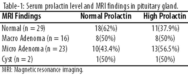
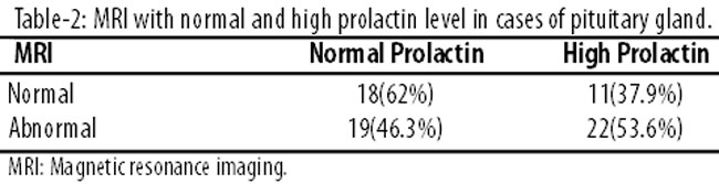
The serum prolactin level in patients with evidence of pituitary microadenoma ranged from 0.7ng/ml to 207ng/ml. In patients with macroadenomas, the level ranged from 0.21ng/ml to 595.7ng/ml. In patients with pituitary cyst, the level ranged from 5.17-26.6ng/ml. The serum prolactin level of patients without evidence of adenoma ranged from 1.1ng/ml to 99.8ng/ml. Hyperprolactinaemia was diagnosed in 32(45.7%) subjects. Out of these, 25 (78.1%) were females and 7 (21.8%) were males. The mean age was 36.4±14.5 years in females (range: 21-75 years) and 43.5±16.4 years (range: 19-69 years) in males. In 25 female patients, 10 (40%) were diagnosed as pituitary microadenomas, 6(24%) were macroadenomas, 8(32%) were normal, and 1(4%) case was diagnosed as pituitary cyst (Table-3).
In 7 male patients, 3(42.8%) were diagnosed as pituitary microadenomas, 1 (14.2%) was macroadenoma, and 3 (42.8%) cases were normal.
Discussion
Pituitary tumours are common neoplasms. Most of our findings are in accordance with literature from the West. Majority of the published literature is in international journals. There is little published literature available in local journals or from Pakistan with focus on serum prolactin level and pituitary tumours.
Pituitary tumours constitute 10-15% of intracranial tumours in surgical specimens.9 Prolactin-secreting tumours are benign neoplasms that account for about 40% of all pituitary tumours. Over 90% are small, intrasellar tumours that rarely increase in size.10 Patients with pituitary tumours present with symptoms of a mass lesion, endocrine dysfunction or both. Endocrine dysfunction may be hyperfunction, hypofunction or both. Prolactinomas are usually seen in clinical practice earlier in females compared to the males; in the latter these are usually macroadenomas.11 The earlier presentation in females is because of the effects of hyperprolactinaemia on gonadal function which is observed early. Serum hormone assay and stimulation/suppression tests are generally sufficient to allow a confident clinical diagnosis of functioning pituitary adenoma. In the low abnormal range (25-200ng/ml), however, many other causes of hyperprolactinaemia are possible, including drugs, metabolic disturbances, chest wall lesions, tumours, sarcoidosis and tuberculosis. It is, therefore, essential to exclude an adenoma.7 Adenoma with a diameter of 3mm seem to be the smallest diameter at which tumours of adenohypophysis can be diagnosed with certainty as microadenomas. Smaller lesions are critical in terms of resolution and could only be vaguely suspected. A previous study12 described a positive correlation between tumour size and serum prolactin level. In a larger series we did not find significant results when correlating serum prolactin levels with tumour size. A microadenoma usually gives lower signal than the surrounding pituitary tissue, before and after contrast medium. Microadenomas rarely enhance. Dynamic MRI of the pituitary with a bolus injection of gadolinium13 demonstrates that adenomas enhance more slowly, peaking later than the normal pituitary tissue.
Prolactinomas are relatively rare in males and more than 70% of the cases occur in females.14 Women with hyperprolactinaemia are usually detected early due to classical presentation of amenorrhoea and galactorrhoea and usually have a microadenoma. The presentation of prolactinoma in males is usually confusing;15 impotence and decreased libido are the most frequent symptoms, but the diagnosis is only made after the signs of compression due to tumour pressure.16 Non-functioning pituitary tumours do not produce any hormones but may cause a deficiency of one or more pituitary hormones by crowding out normal pituitary tissue or prolactin excess by causing stalk compression. Of our subjects, 22.8% were diagnosed to have non-functioning pituitary tumours. Approximately 25-30% of patients are described to have non-functioning pituitary tumours in the literature.17
Some authors have reported a positive correlation between tumour volume and serum prolactin value.18 This might be true for patients with hyperprolactinaemia. However, when patients without hyperprolactinaemia are also included as in the present study, where some of the patients had normal serum prolactin values, many of them with large tumours, no such correlation is found.
A small sample size and being a single-centre study, the current study does have its limitations. In addition, the study shares the limitations of all retrospective studies.
Conclusion
MRI pituitary findings were not associated with serum prolactin levels in patients with clinical suspicion of pituitary abnormality. Therefore, regular monitoring of serum prolactin is suggested. Further large multi-centre studies are suggested to establish the association.
References
1. Kulkarni MV, Lee KF, McArdle CB, Yeakley JW, Haar FL. 1.5-T MR imaging of pituitary microadenomas: technical considerations and CT correlation. Am J Neuroradiol 1988; 9:5-11.
2. Elster AD. Modern imaging of the pituitary. Radiology 1993; 187:1-14.
3. Bonneville JF, Cattin F, Dietemann JL. Computed tomography of the pituitary gland. New York: Springer-Verlag, 1986; pp 63-88.
4. Bilaniuk LT, Zimmerman RA, Wehrli FW, Snyder PJ, Goldberg HI, Grossman RI, et al. Magnetic resonance imaging of pituitary lesions using 1.0 to 1.5 T field strength. Radiology 1984; 153:415-8.
5. Pojunas KW, Daniels DL, Williams AL, Haughton VM. MR imaging of prolactin-secreting microadenomas. Am J Neuroradiol 1986; 7:209-13.
6. Rand T, Kink E, Sator M, Schneider B, Huber J, Imhof H, et al. MRI of microadenomas in patients with hyperprolactinaemia. Neuroradiology 1996; 38:744-6.
7. Peck WW, Dillon WP, Norman D, Newton TH, Wilson CB. High-resolution MR imaging of microadenomas at 1.5 T: experience with Cushing disease. Am J Roentgenol 1989; 152:145-51.
8. Lundin P, Pedersen F. Volume of pituitary macroadenomas: assessment by MRI. J Comput Assist Tomogr 1992; 16:519-28.
9. Kovacs K, Horvath E. Pathology of pituitary tumors. Endocrinol Metab Clin North Am 1987; 16:529-551.
10. March CM, Kletzky OA, Davajan V, Teal J, Weiss M, Apuzzo ML, et al. Longitudinal evaluation of patients with untreated prolactin-secreting pituitary adenomas. Am J Obstet Gynecol 1981; 139:835-44.
11. Lamberts SW, de Herder WW, Kwekkeboom DJ, vd Lely AJ, Nobels FR, Krenning EP. Current tools in the diagnosis of pituitary tumours. Acta Endocrinol 1993; 129 suppl 1: 6-12.
12. Lundin P, Nyman R, Burman P, Lundberg PO, Muhr C. MRI of pituitary macroadenomas with reference to hormonal activity. Neuroradiology 1992; 34:43-51.
13. Miki Y, Matsuo M, Nishizawa S, Kuroda Y, Keyaki A, Makita Y, et al. Pituitary adenomas and normal pituitary tissue: enhancement patterns on gadopentetate-enhanced MR imaging. Radiology 1990; 177:35-8.
14. Nabarro JDN. Review: pituitary prolactinomas. Clin Endocrinol 1982; 17:129-55.
15. Jackson JA, Kleerekoper M, Parfitt AM. Symptomatic osteoporosis in a man with hyperprolactinemic hypogonadism. Ann Intern Med 1986; 105:543-45.
16. Delgrange E, Trouillas J, Maiter D, Donckier J, Tourniaire J. Sex-related difference in the growth of prolactinomas: a clinical and proliferation marker study. J Clin Endocrinol Metab 1997; 82:2102-7.
17. Hulting AL, Muhr C, Lundberg PO, Werner S. Prolactinomas in men: clinical characteristics and effect of bromocriptine treatment. Acta Med Scand 1985; 217:101-9.
18. Klijn JG, Lamberts SW, de Jong FH, Docter R, van Dongen KJ, Birkenhäger JC. The importance of pituitary tumour size in patients with hyperprolactinaemia in relation to hormonal variables and extrasellar extension of tumour. Clin Endocrinol 1980; 12:341-55.
Related Articles
Journal of the Pakistan Medical Association has agreed to receive and publish manuscripts in accordance with the principles of the following committees:


