

Qurban Hussain ( Medical Unit IV, Medical ICU, Jinnah Postgraduate Medial Centre, Karachi. )
Shahbaz Haider ( Medical Unit IV, Medical ICU, Jinnah Postgraduate Medial Centre, Karachi. )
Noor Muhammad Solangi ( Medical Unit IV, Medical ICU, Jinnah Postgraduate Medial Centre, Karachi. )
Liaquat Ali ( Medical Unit IV, Medical ICU, Jinnah Postgraduate Medial Centre, Karachi. )
Hammad Liaquat ( Medical Unit IV, Medical ICU, Jinnah Postgraduate Medial Centre, Karachi. )
Fayyaz Ahmed ( Department of Radiology, Jinnah Postgraduate Medial Centre, Karachi. )
Sumera Shahbaz ( Department of Radiology, Jinnah Postgraduate Medial Centre, Karachi. )
May 2013, Volume 63, Issue 5
Original Article

Abstract
Objective: To observe the changes in doppler waveform of hepatic vein after the administration of terlipressin, and to assess indirectly the efficacy of the drug to reduce the Hepatic Vein Pressure Gradient and portal pressure.
Methods: The quasi-experimental study was conducted at the Jinnah Postgraduate Medical Centre, Karachi, from April 1 to November 25, 2011, and comprised 50 patients with cirrhosis with abnormal doppler waveform of the hepatic vein. Patients with diseases causing abnormal hepatic vein doppler waveform were excluded. Doppler waveforms were studied for 20 minutes before and for 20 minutes after the administration of terlipressin. Tracings with best waveform before and after injection were saved for analysis. Changes in waveform after vasoactive drug were defined as mild, significant, marked and gross changes. SPSS 10 was used for statistical analysis.
Results: Of the 50 patients, 36 (72%) were males and 14 (28%) females. Commonest waveform was monophasic 38(76%). Gross changes i.e. turning triphasic from monophasic waveform was observed in 8 (16%) patients. Significant gross changes were seen in 24 (48%) patients. Total number of patients showing improvement in waveform was 36 (72%). In no case, waveform deteriorated after the administration of terlipressin (p= 0.001).
Conclusion: Non-invasive method of observing the improvement of hepatic vein waveform by duplex ultrasound, after more studies, may be an important tool for assessing and monitoring the effects of portal pressure lowering drugs.
Keywords: Doppler waveform, Portal hypertension, Terlipressin, Hepatic vein, Cirrhosis. (JPMA 63: 604; 2013).
Introduction
Decompensated liver cirrhosis, final or end-stage liver disease, is characterised by the presence of either one or more of ascites, hepatorenal syndrome, variceal haemorrhage and hepatic encephalopathy.1-3
Portal hypertension, important precursor of variceal haemorrhage, is caused by deficient intra-hepatic vasodilator nitric oxide, increased intra-hepatic vasoconstrictors like endothelin, angiotensin, nor-epinephrine and cysteinyl-leukotrienes, as well as by intra-hepatic fibrosis, a well-known and most important feature of cirrhosis.4-7 Portal hypertension and drugs lowering the portal hypertension have been the focus of interest for a number of decades, as portal hypertension leads to life-threatening variceal bleed. To measure pressure in the portal vein is very difficult technically as portal vein, after receiving blood from the stomach and gut capillaries, in liver divides again and again into smaller and smaller vessels to supply the hepatocytes.8 Hepatic vein (HV), its free pressure and pressure while catheter is in wedged position, have also been studied to assess the severity of portal hypertension in decompensated cirrhosis.9
It has been shown that Hepatic Vein Pressure Gradient (HVPG), which is calculated after subtracting free hepatic venous pressure from the wedged hepatic venous pressure, is a good indicator of portal hypertension. HVPG calculating formula is as follows:10-12
HVPG= Wedged hepatic venous pressure - Free hepatic venous pressure.
An HVPG more than 15 mmHg is defined as severe hypertension.11-13 Continuing studies on hepatic vein led to focus on doppler hepatic vein waveform and then on comparison of doppler waveform of hepatic vein and HVPG14-18 This comparison showed that HVPG is closely related to doppler hepatic vein waveform which are labelled classically as \\\'triphasic\\\', \\\'biphasic\\\' and \\\'monophasic\\\'.19 \\\'Triphasic\\\' waveform of hepatic vein is seen in normal subjects with normal liver, normal heart and normal portal vein pressure.19-21 With increasing portal hypertension, HV waveform is described to change to \\\'biphasic\\\' and then to \\\'monophasic\\\' waveform.21-23 Comparison of HVPG and doppler waveform of HV has shown that shift in doppler waveform of hepatic vein from normal and increase in HVPG is closely related.12 Changes in waveform from \\\'monophasic\\\' to \\\'biphasic\\\'/\\\'triphasic\\\' or from \\\'biphasic\\\' to \\\'triphasic\\\' after administration of certain vasoactive drugs (portal pressure lowering drugs) have been found to be closely and reliably associated with decrease in HVPG.11,12,22 This observation is very important for studying the effects of certain portal pressure lowering drugs because invasive methods e.g. HVPG may be replaced by the non-invasive doppler waveforms studies before and after the administration of the drugs. In one study12 change of waveform from \\\'monophasic\\\' to \\\'biphasic\\\' , from \\\'monophasic\\\' to \\\' triphasic\\\' and from \\\'biphasic\\\' to \\\'triphasic\\\' correlated with decrease in HVPG 3-10 mm of Hg (mean 5.8), 5-9 (mean 7) and 5-9 (mean 7) mm of Hg respectively. The change observed with waveform change was always 3 or more than 3mmHg.
The knowledge obtained from similar studies has been used for planning of this study.12,22 The objective of this study was to observe doppler waveform of hepatic vein before and after the administration of injection terlipressin 2mg and thus to observe indirectly, by change in waveform, its efficacy to reduce the HVPG and portal hypertension. It also tried to assess the non-invasive doppler waveform investigation as a monitoring tool for changes in portal pressure.
Patients and Methods
The quasi-experimental study was carried out after approval by Ethical Committee of Jinnah Postgraduate Medical Centre (JPMC) from April 1 to November 25, 2011 when the required number of 50 cases was achieved. Written informed consent was taken from all the patients who were selected from among those attending Out-patient Department of Medical Unit-IV, patients admitted to the unit and to the Medical intensive care unit (ICU) of JPMC.
Diagnosis of liver cirrhosis was established by clinical examination, lab data, imaging studies, including ultrasonography, computed tomography scanning, ascitic fluid examination and upper gastrointestinal (GI) Endoscopy. Patients who did not provide informed consent or had hepatocellular carcinoma, hepatic encephalopathy, severe liver failure (serum bilirubin >5mg/dl {>85µm/l}), thrombus in the hepatic/portal vein or inferior vena cava, congestive cardiac failure or any other cardiac disease causing dilated inferior vena cava were excluded from the study. Patients having \\\'triphasic\\\' waveform on initial Doppler waveform study and patients unstable haemodynalmically were also excluded. No patient had been taking any drug that affects haemodynamics for the preceding 10 days.
Patients were selected and their doppler waveform study was done by an experienced radiologist. The GE Model Volusion ultrasound machine was used. Convex probe was placed intercostally at 190Hz.
Initially, the hepatic vein was focused, then doppler shift signals were obtained from the right hepatic vein at a distance of 3-4cm from the junction of the hepatic vein and inferior vena cava.
HV doppler waveforms were observed for 20 minutes before the injection, and the whole process of observing and recording doppler waveforms was repeated for 20 minutes after intravenous bolus injection of 2mg of terlipressin (Novapressin TM). Waveforms were recorded and tracings with best waveform during the two observation periods i.e. before and after the injection as described were saved for analysis. Each tracing was recorded for 5-6 seconds with end expiration breath holding. These tracings recorded were labelled as \\\'monophasic\\\' to \\\'triphasic\\\' waveforms. While labelling doppler waveforms intermediate classes like \\\'mono-to-biphasic\\\' and \\\'bi-to-triphasic\\\' waveform were also used in addition to classic \\\'triphasic\\\' . \\\'biphasic\\\' and \\\'monophasic\\\' waveforms. These waveforms were defined as: \\\'Monophasic\\\': Flat line or wavy (with no phasic change in amplitude of wave); \\\'Mono-to-biphasic\\\': Wavy with phasic change in amplitude but change not regular; \\\'Biphasic\\\': Regular phasic change in amplitude of waves, but no reversal of flow (i.e. no 3rd phase); \\\'Bi-to-triphasic\\\': Regular biphasic oscillation with slight and irregular flow reversal wave; and \\\'Triphasic\\\': Regular phasic oscillation with flow reversal wave.
Study continued till the 50 patients were observed for the effects of single dose of terlipressin. Age group and gender of the patients were analysed statistically. Mean age and standard deviation were determined. Determining the category of waveform out of these five \\\'monophasic\\\' to \\\'triphasic\\\' categories was done by two radiologists. These categories were quantified by assigning 1 to \\\'monophasic\\\'; 2 to \\\'mono-to-biphasic\\\'; 3 to \\\'biphasic\\\'; 4 to \\\'bi-to-triphasic\\\'; and 5 to \\\'triphasic\\\' waveform. The quantified change after injection terlipressin 2mg was calculated by subtracting the number of later waveform from the number of initial waveform. For instance, if after injection terlipressin, the waveform was found to be changed from \\\'mono-to-biphasic\\\' (assigned number 2) to \\\'triphasic\\\' (assigned number 5), the change was determined as 5 - 2 i.e. 3. This change was then defined as \\\'mild\\\' if result of subtraction was 1; \\\'significant\\\' for result of 2; \\\'marked\\\' change for a subtraction result of 3; and \\\'gross\\\' change for a subtraction result of 4.
Statistical analysis was performed on SPSS 10, while \\\'p\\\' value was determined by applying Wilcoxon matched pair test.
Results
Of the 50 patients enrolled, 36 (72%) were males and 14 (28%) were females. Maximum number (n=17; 34%) belonged to the age group 40-49 years followed by 10 (20%) in age groups of 50-59 and 60-69 years each. The mean age was 44.66±13.34 years.
Direction of blood flow in the portal vein was observed. It was hepatopetal in 46 (92%) and hepatofugal in 4(8%) patients. Mean blood velocity in the hepatic vein was 27.17±9.55cm/sec. After injection terlipressin, hepatic vein velocity increased in 36 (72%) patients and decreased in 14 (28%).
Regarding initial doppler waveform of the hepatic vein, commonest was \\\'monophasic\\\' pattern i.e. in 38 (76 %). Patients having \\\'triphasic\\\' doppler waveform were excluded from the study , as no improvement in waveform was possible after the adminstration of terlipressin (Table-1).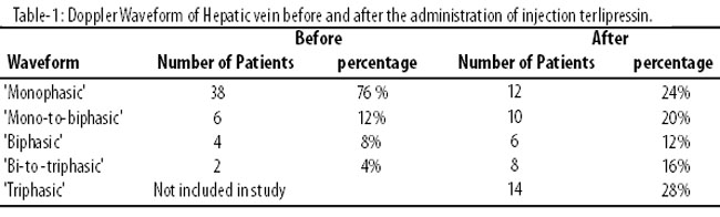
Quantified change in wave-form after the injection was calculated. No change was observed in 14 (28%) cases, while in 36(72%) waveform improved and improvement ranged from \\\'mild\\\' to \\\'gross\\\' (Table-2).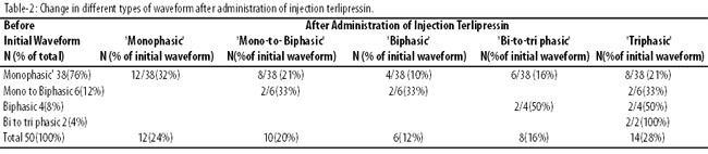
Discussion
To decrease pressure in the portal vein is the basic aim to prevent or stop variceal bleeding in patients of liver cirrhosis. Improvement in doppler waveform like lowering of HVPG, is an important detector of change in portal pressure. A study looked into doppler waveforms before and after the administration of 2mg of Terlipressin.12 Out of 12 patients having \\\'monophasic\\\' waveform before the administration of terlipressin, 9 (75%) patients had their doppler waveforms turned to \\\'biphasic\\\'. In the other 3 (25%) patients, the waveform improved even more and turned to \\\'triphasic\\\'. Among the 8 patients having \\\'biphasic\\\' waveform, 6 (75%) patients had it improved to \\\'triphasic\\\', and in 2 (25%) the waveform remained unchanged. In that study12 terminology of intermediate forms i.e. \\\'mono-to-biphasic\\\' or \\\'bi-to-triphasic\\\', was not used. However, to compare this similar but not identical study12 with our effort (Table-3),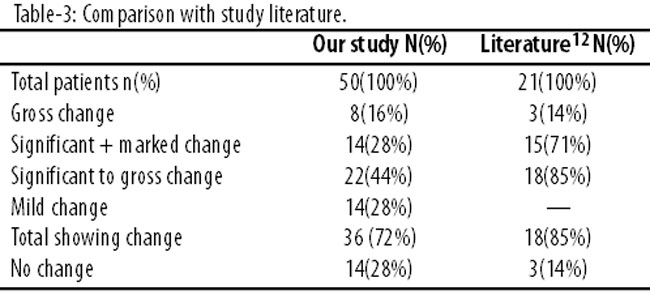
we quantified the results in a method similar to how we went about it.
In our study waveforms were labelled more precisely using \\\'mono-to-biphasic\\\' and \\\'bi-to-triphasic\\\' in addition to \\\'monophasic\\\' , \\\'biphasic\\\' and \\\'triphasic\\\'. Furthermore , we did not include patients with initial \\\'triphasic\\\' waveform as was done in that study,12 as no improvement in waveform was expected. Percentage of cases of improvement in waveform were lower in our study than those of the other.12 As improvement in doppler waveform is an indicator of decrease in HVPG , it shows, like decrease in HVPG, a decrease in portal hypertension. This decrease in HVPG was also shown by a study24 in which significant decrease in HVPG was observed along with decrease in portal venous blood flow after the injection. In our study, portal vein blood velocity decreased in 30 (60%) cases and increased in 20 (40%).
Importance of observation of positive changes in the hepatic vein waveforms as seen by doppler ultrasound lies in the fact that it is non-invasively observed; cost per patient observation is negligible compared to invasive HVPG measurement; procedure can be repeated easily; and effects of multiple drugs may be observed at appropriate intervals.
Abnormalities of waveforms of the hepatic vein have not been easy to explain.12 Change of hepatic vein compliance in cirrhosis had been suggested as explanation of abnormal hepatic vein doppler waveform. But terlipressin-induced changes in waveform suggest that a haemodynamic effect of high portal pressure rather than a fixed structural abnormality is the pathogenic mechanism responsible for the abnormal waveforms.12 Structural changes in liver parenchyma leading to fibrosis and obstruction to portal flow passing through the liver is widely accepted mechanism leading to portal hypertension and gastroesophageal varices.1,2 But, as expected, this should lead to decrease in the hepatic blood flow as considerable quantity of portal blood is going towards other systemic veins via portosystemic anastomosis instead of hepatic veins.2 Terlipressin, a synthetic analogue of vasopressin (a vasoconstrictor agent), is said to cause vasoconstriction of splanchnic arteries.24 This vasoconstriction results in reduced venous drainage from stomach and intestine, thus reducing portal venous flow and portal vein pressure within minutes.24 By reducing portal blood flow through liver, terlipressin reduces the already reduced hepatic vein blood flow. Doppler waveform of hepatic vein in cirrhosis turns abnormal in which major change is reduction of blood flow through hepatic vein due to fibrotic changes affecting both portal and hepatic artery blood supply. It is difficult to answer why the further reduction of blood flow by the vasoconstrictor drugs like terlipressin shifts the waveform towards normal. These questions require more insight into the issue. One explanation given in literature is that "high portal pressure probably contributing to the flattening of the normal \\\'triphasic\\\' hepatic waveform haemodynamically blunting the effect of variation in central venous pressure during cardiac cycle."12 But keeping the anatomy8 in mind, question arises, how the high pressure in the portal vein , which is maximum near porta hepatis and would have lowered down due to fibrotic obstructions before and after sinusoidal capillaries, may assert its effects on anatomically and functionally distantly located hepatic veins which are directly draining into the inferior vena cava?
Conclusion
In liver cirrhosis, doppler waveform abnormalities of the hepatic vein were seen. After the administration of injection of vasoactive agent, terlipressin, most of the cases showed improvement in waveform. As these improvements showed decrease in HVPG and portal pressure, non-invasive doppler waveform studies, in future, are expected to be an important tool for assessing and monitoring the effects of portal pressure lowering drugs. However, to understand how does waveform improve in anatomically and functionally distantly located hepatic veins, more research on portal and hepatic vein flow patterns is required.
References
1. Lawrence S, Friedman .Liver , Biliary tract and Pancreas Disorder. In: Papodakis M, McPhee SJ, Robow MW. Current Medical Diagnosis and Treatment. 49th ed. USA: McGraw Hill comparis. 2010; pp 644-98.
2. Lebrec D, Vinel JP, Dupas JL .Complications of portal hypertension in adults: a French consensus Eur J of Gastroenterol and Hepatol 2005; 17: 403-10.
3. Spahr L, Giostra E, Frossard JL, Morard I, Mentha G, Hadengue A.A 3-month course of long-acting repeatable octreotide (sandostatin LAR) improves portal hypertension in patients with cirrhosis: a randomized controlled study. Am J Gastroenterol 2007; 102: 1397-405.
4. Lin HC, Yang YY, Hou MC, Huang YT, Lee FY, Lee SD. Acute administration of carvedilol is more effective than propranolol plusisosorbide-5-mononitrate in the reduction of portal pressure in patients with viral cirrhosis. Am J Gastroenterol 2004; 99: 1953-8.
5. Wallace JL. The vascular endothelium and nitric oxide. In: Groszmann RJ, Bosch J. Portal Hypertension in 21st Century. (ebook) ISBN 978-94-010-3774-7. 2004; pp 13-4.
6. Amico G, Garcia-Pagan JC, Luca A, Bosch J. Hepatic vein pressure gradient reduction and prevention of variceal bleeding in cirrhosis: a systematic review. Gastroenterology 2006; 131: 1611-24.
7. Carlos J. Enhanced release of vasoconstrictors. In: Portal Hypertension. In: Groszmann RJ, Bosch J. 21st century. (ebook) ISBN 978-94-010-3774-7. 2004; pp 89-96.
8. Ryan SP, McNichdes MMJ, Eustace SJ. Anatomy for Diagnostic Imaging. 2nd ed. Philadelphia: Saunders; 2004; pp 187-9.
9. Groszmann RJ, Wongcharatrawee S. The hepatic venous pressure gradient: anything worth doing should be done right. Hepatology 2004; 39: 280-2.
10. Huet PM, Pomier-Layrargues G. The hepatic venous pressure gradient: "remixed and revisited." Hepatology 2004; 39: 295-8.
11. Vorobioff JD, Gamen M , Kravetz D, Picabea E, Villavicenio R, Bordata J, et al. Effects of long-term propranolol and octreotide on postprandial hemodynamics in cirrhosis: a randomized, controlled trial. Gastroenterology 2002; 122: 916-22.
12. Baik SK, Kim JW, Kim HS, Kwon SO, Kim YJ, Park JW, et al. Recent variceal bleeding: Doppler US hepatic vein waveform in assessment of severity of portal hypertension and vasoactive drug response. Radiology 2006; 240: 574-80.
13. Hussain Q, Badruddin AH, Chaudhry MA, Ahmad F, Abbasi A. Effect of carvedilol on portal pressure estimated by hepatic vein Doppler ultrasound waveform and damping index in cirrhotic patients. J Coll Physicians Surg Pak 2010; 20: 586-9.
14. Baik SK, Jee MG, Jeong PH, Kim JW, Ji SW, Kim HS, et al. Relationship of hemodynamic indices and prognosis in patients with liver cirrhosis. Korean J Intern Med 2004; 19: 165-70.
15. Sato S, Ohnishi K, Sugita S, Okuda K. Splenic artery and superior mesenteric artery blood flow: nonsurgical Doppler US measurement in healthy subjects and patients with chronic liver disease. Radiology 1987; 164: 347-52.
16. Choi YJ, Baik SK, Park DH, Kim MY, Kim HS, Lee DK. Comparison of Doppler ultrasonography and the hepatic venous pressure gradient in assessing portal hypertension in liver cirrhosis. J Gastroenterol Hepatol 2003; 18: 424-9.
17. Merkel C, Sacerdoti D, Bolognesi M, Bombonato G, Gatta A. Doppler sonography and hepatic vein catheterization in portal hypertension: assessment of agreement in evaluating severity and response to treatment. J Hepatol 1998; 28: 622-30.
18. Taourel P, Blanc P, Dauzat M, Chabre M, Pradel J, Gallix B, et al. Doppler study of mesenteric, hepatic, and portal circulation in alcoholic cirrhosis: relationship between quantitative Doppler measurements and the severity of portal hypertension and hepatic failure. Hepatology 1998; 28: 932-6.
19. Ghazaleh Tabatabai, Hoffer M. Abdomen. In: Hofer M (editor) Teaching manual of color duplex sonography: a workbook on color duplex. 2nd ed. New York: Georg Thieme Verlag; 2010; pp 39-40.
20. Coulden RA, Lomas DJ, Farman P, Britton PD. Doppler ultrasound of the hepatic veins: normal appearances. Clin Radiol 1992; 45: 223-7.
21. Ethan J, Halpern ET. Sience to practice: noninvasive assessment of portal hypertension - can US aid in the prediction of portal pressure and monitoring of therapy? Radiology 2006; 240: 309-10.
22. Kim MY, Baik SK, Park DH, Lim DW, Kim JW, Kim HS, et al. Damping index of Doppler hepatic vein waveform to assess severity of portal hypertension and response to propranolol in liver cirrhosis: a prospective non-randomized study. Liver Int 2007; 27: 1103-10.
23. Bolondi L, Bassi SL, Gaiani S, Zironi G, Benzi G, Santi Y, et al. Liver cirrhosis: changes of Doppler waveform of hepatic veins. Radiology 1991; 178: 513-6.
24. Baik SK, Jeong PH, Sang WJ,Yoo BS, Kim HS , Lee DK, et al. Acute hemodynamic effects of octreotide and terlipressin in patients with cirrhosis: a randomized comparison. Am J Gastroenterol 2005; 100: 631-5.
Related Articles
Journal of the Pakistan Medical Association has agreed to receive and publish manuscripts in accordance with the principles of the following committees:


