

Asif Khalid ( Department of Oncology, Allama Iqbal Medical College, Services Hospital, Lahore. )
Maliha Zahid ( Department of Oncology, Allama Iqbal Medical College, Services Hospital, Lahore. )
Amjad Rehman ( Department of Oncology, Allama Iqbal Medical College, Services Hospital, Lahore. )
Zaheer-Ud-Din Ahnmd ( Department of Oncology, Allama Iqbal Medical College, Services Hospital, Lahore. )
Sobia Qazi ( Department of Oncology, Allama Iqbal Medical College, Services Hospital, Lahore. )
Zeba Aziz ( Department of Oncology, Allama Iqbal Medical College, Services Hospital, Lahore. )
April 1997, Volume 47, Issue 4
Original Article

Abstract
A total of 113 patients of leukemia, over 15 years of age, were seen in three different institutions from July, 1992 to June, 1994. There was an almost equal distribution of acute myeloid leukemia (AML) and acute lymphoblastic leukemia (ALL) (44 vs 43 cases respectively). Chronic lymphocytic leukemia (CLI.) was the least common, accounting for 5% of all cases. Mean age in CLL was 59 yeats. Chronic myeloid leukemia (CML) was three times commoner than CLL with a younger age distribution (median age was 34 years). We conclude that the clinicoepidemiological features of adult leukemias differ considerably from that seen in the developed world. However, recruitment of patients needs to continue in order to define these features based on a larger patient population (JPMA 47:119, 1997).
Introduction
Acute and chronic adult leukemias are a heterogenous group of disorders. They are also an uncommon form of malignancy affecting approximately 5 persons per 100,000 in the United States annually1. The causes remain largely unknown and hypothetical. Epidemiological studies concentrate on small variabilities in the incidence seen in different countries and within countries2,3. Etiological factors in development of leukemias are hereditary disorders with susceptibility to chromosomal breakage4,5 exposure to radiation6,7 and chemicals like benzene8-11. This article focuses on basic clinicoepidemiological features of adult leukemias in Pakistan. It highlights some important differences in distribution of subtypes of leukeniias, age at presentation and presenting signs and symptoms as compared to those from North America.
Patients and Methods
From July, 1992 to June, 1994 a total of 171 patients of Leukemia presented to the Department of Oncology. The diagnosis was confinned on bone marrow aspiration and trephine biopsy. Of these, 113 patients were above 15 years of age, 58 belonged to the pediatric age group and were excluded from the analysis. A precoded proforma for collecting data on the epidemiology of leukemias was developed and filled forall patients. The factors included age, sex, subtypes of disease, socioeconomic status and ethnic group. Prior histoiy of exposure to chemicals and radiation was sought. Family history for hematological malignancies, solid tumors and non-malignant disorders was obtained. Presenting signs, symptoms and performance status of the patients was recorded. The data was analysed using database management system.
Results
Of 113 cases, 43 (3 8%) were of acute lymphoblastic, 44 (39%) acute myeloid and 20 (18%) chronic myeloid leukemias. There were only 6 (5%) cases of chronic lymphocytic leukemia.
Acute lymphoblastic leukemia (ALL)
Forty-three cases of ALL were seen. There was significant male predominance. The presenting ages ranged from 16 to 94 years (mean of 33 years). The mean duration of symptoms was 6 months (Table I, Figure 1).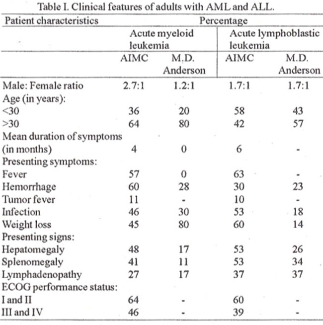
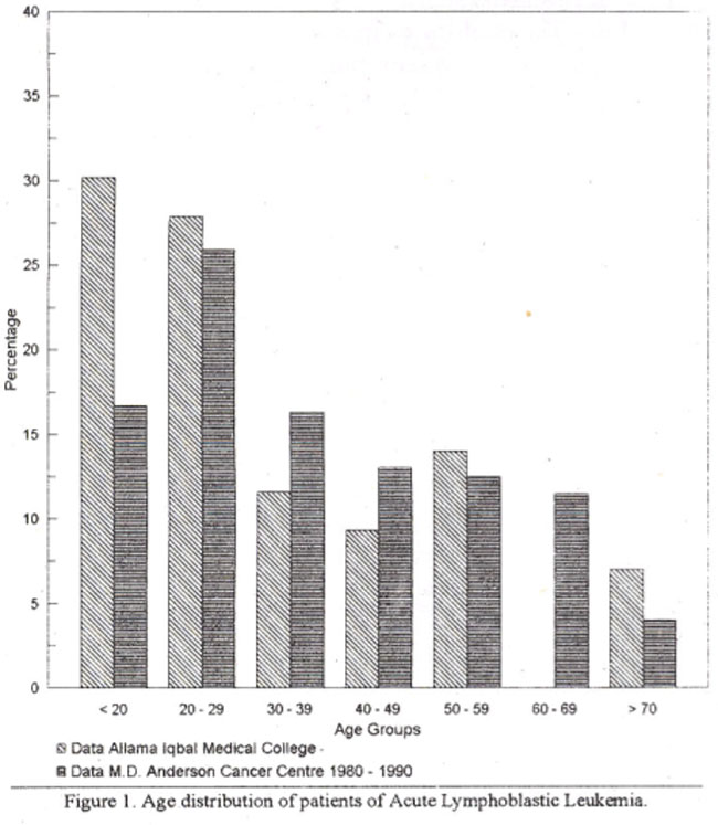
Majority of patients were Punjabis (93%). belonging to middle class (56%). Exposure to pesticides and tobacco was infrequentand only 5% of cases used alcohol. No patient reported exposure to radiation (Table II). The patients were generally a healthy population pnorto developing leukemia, only one had Downs syndrome. Positive family history of leukemiawas obtained in one and hepatocellular carcinoma in another patient. No familial or geographical clustering of leukemias was identified. Two patients had a family history of ischemic heart disease, diabetes mellitus and renal calculi (Table II).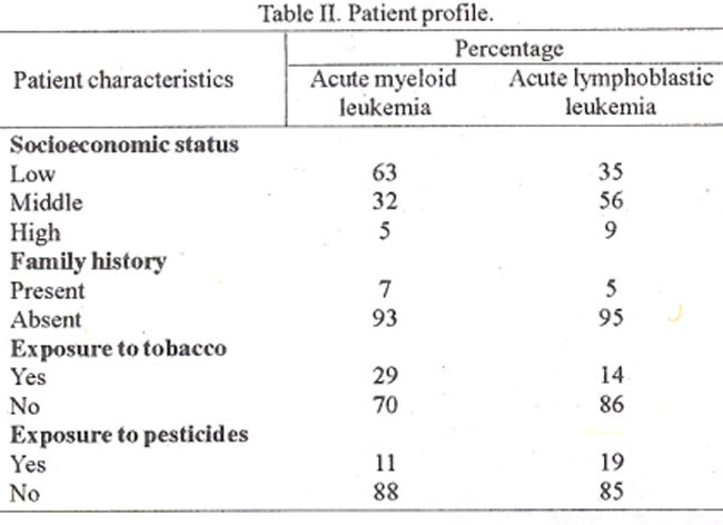
ECOG (Eastern Cooperative Oncology Group) performance status land II was present in 60% and status III orIV in 39% of cases. The main presenting symptoms were fever, weight loss and bleeding and signs hepatosplenomegaly and lymphadenopathy (Table I). Tumor fever was present in 84% patients and 16% had documented infections. Only three patients had received priorchemothenpy. Majority of patients presented immediately after diagnosis.
Acute Myeloid Leukemia (AMIL)
A total of 44 patients of AML were seen. Their ages ranged from 17 to 72 years ( median 36 years)as shown in Figure 2.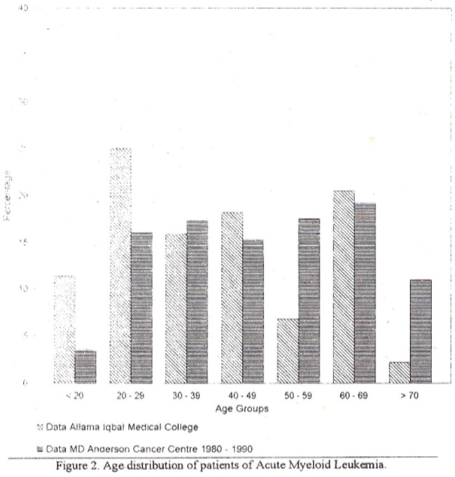
The avenge duration of symptoms was 4 months. Male to female ratio was 2.7:1. Majority were Punjabis who belonged to the middle socioeconomic class. History of exposure to chemicals like fertilizers, insecticides and pesticides was present in 11.5% cases. Only one patient gave a history of exposure to radiation. There were 29% smokers while only one gave a history of alcohol use (Table II). Majority (84%) were healthy prior to developing acute leukemia. Coronary artery disease and diabetes mellitus was present in 16% of patients and none had a prior history of cancer. Family history of cancer was present in 7% cases (Table II). There was one case each of breast cancer and two cases of hepatocellular carcinoma. Using WHO perfonnance status 64% belonged to land 11,36% had a performance status of ill or IV The commonest complaint was bleeding/purpura followed by fever and weakness/weight loss. On presentation 59% were febrile of which 81% had tumor fever while infections accounted for 19%. Bleeding diathesis was present in 64% cases, 48% had hepatomegaly, 41% splenomegaly and 27% lymphadenopathy. Sixteen percent received chemotherapy and 2% had undergone radiation therapy prior to presentation.
Chronic myeloid leukemia (CML)
Chronic myeloid leukemia was diagnosed in 20 patients. The age ranged from 16 to 55 years (median 34 years). Male to female ratio was 1.75:1. The mean duration of symptoms was 19 months. More than 90% of the patients were Punjabis. The population was evenly divided between the lower and middle socioeconomic group. Nearly 60% were unemployed, 10% had history of exposure to insecticides or pesticides, 10% were smokers and none gave history of exposure to radiation. Three (15%) patients reportedapositive family history of hypertension, ischemic heart disease or chronic respiratory diseases. Majority (82%) of the patients belonged to the WHO performance status I and II and 18% to performance status III. The commonest complaints were weakness/malaise (40%), followed by bleeding/purpura (30%), fever (30%) and cough (10%); 27% had fever on presentation. The fever was due to the underlying malignancy in 89% of the cases with infections accounting for 11% of febrile episodes. On presentation 90% of the patients had splenomegaly, 60% had lymphadenopathy and 50% had hepatomegaly. Fourpatients had received pnor chemotherapy with Myleran and one had received radiation therapy to the spleen.
Chronic lymphocy tic leukemia (CLL)
CLL was the least common leukemia, accounting for only six outof 113 patients. The age rangedfrom45 to 65 years (average age 59 years). The male to female ratio was 2:1. The mean duration of symptoms were 12 months. Five patients were from the lower socioeconomic group and one belonged to the middle class. Two cases reported exposure to pesticides and fertilizers. No patient gave a history of smoking oralcohol use. There was no history of exposure to radiation. All cases with CLL reported are well. Family history of hepatocellular cancer and tuberculosis was present in one patient each. There were four patients with performance status group I and II and two with ECOG performance status III and IV. The chief complaints were fever (3 cases), weakness/weight loss (2 cases), bleeding/purpura (1 case) and cough (1 case). Two patients had infectious fever on presentation, 2 had a bleeding diathesis, three had hepatomegaly. five had splenomegaly and four lymphadenopathy.
Discussion
Hematological malignancies, especially leukemias and non- Hodgkin’s lymphoma are common in South-East Asia than in the U.S.A. and Europe12. In a study, based on tumor registry data by Armed Forces Institute of Pathology, Leukernias were the second most common malignancy seen in the adult male patient population and the fourth commonest malignancy in the adult female patient population from Northern Pakistan13. Data from Karachi, reported leukemias to be the fifth commonest malignancy in adult patients of both sexes14. This is in contrast to the data based on NCI SEER Program (National Cancer institute and Surveillance, Epidemiology and End Results) (1985-1987) which reported adult leukernias to be an uncommon fonn of malignancy affecting approximately 5 persons per 100,000 in the United States annually1.
Acute myeloid leukemia, is five times more common than ALL’. In China AML is three times more common than ALL12,15. AML and ALL had a similar distribution in our study wth a male predominance similar to that reported in literature16. AMIL is more common in patients older than 50 years of age’. In our patients peak incidence occurred in the third decade of life with no progressive increase in incidence with increasing age. Similar observations have been reported by others17 who showed a peak incidence between 31 to 40 years in 54 patients of AML. Hassan et al18 reported 62 cases of AML where all morphological subtypes (except M4) occurred in ages between 25 and 29 years. The mean age of patients with M4 disease was 46 years. Acute lymphocytic leukemia (ALL) on the other hand showed peaks in the second and third decade of life. Leukemia are more common in higher socioeconomic settings19, however majority of our patients belonged to low or middle class; only 10% belonged to the higher socioeconomic group. Since these patients are from three different institutions, a wide socioeconomic spectrum was covered and the difference is probably not due to selection bias alone. The etiology of most leukeniias, especially adult ALL, are unknown. The contribution of heredity to leukemia has become increasingly important because of frequent genetic reanangements. The strongest association of heredity is seen with Downs syndrome, in which there is a 20 fold increased risk of leukemia4. Hereditary disorders with a tendency to chromosomal breakage are associated with an increased risk of AML5. Chrornosornal abnormalities were reported from AFIP20, Rawalpindi, in 57% of 35 cases studied. Exposure to radiation, therapeutically or accidently, leads to an increased risk of acute leukemias as seen in the survivors of the atomic bomb explosions in Japan6,7. The overall incidence of leukemia increased 10 to 15 fold and was greater for ALL then AML. The association of therapeutic irradiation with leukemia is best demonstrated in patients with ankylosing spondylitis in whom radiation to the spine was associated with a five fold increase in acute leukemia2. There was no exposure to radiation in our patients. Increased risk of AML with cigarette smoking in certain cytogenetic subgroups2’ has been reported. Studies seeking an association with the use of pesticides by farmers have not provided strong evidence for increased risk22,23. In the present study 30% patients gave a history of exposure to pesticides and fertilizers, however an association can only be proved by long- term case-control studies. The mean duration of symptoms before presentation for ALL and AML were 6 and 4 months respectively, this is in contrast to the data reported in Western literature where patients present earlier. As a result of this delayed presentation, a higher proportion of patients had bleeding diathesis, weight loss, hepatomegaly, splenomegaly and lymphadenopathy. Similar findings have also been reported by others from Pakistan. Hepatomegaly and splenomegaly were uncommon inAML. Palpable lymph node enlargement was also more common in patients with ALL. As reported in literature symptoms of petechiae and ecchyrnoses were more common in our patients of AMIL.
CLL is the commonest leukemia of all, with an annual incidence of 1.8 to 3.0 per 100,000 population, in the United States. Incidence is age related and increases with increasing age. It affects twice as many men as women. In contrast it was the least common of all leukemias seen in our patients with a significant male predominance. This decreased incidence of CLL has also been observed in Japanese and other Asian populations. The relatively younger patient population, with a mean of 59 years, were affected probably due to shorter life expectancy in our country. Chronic myeloid leukemia in our populationwas three times as common as CLL. It also affected a younger age group with an age range of 16 to 55 years. Eight outof20 patients were under3 Oyears ofage. Thisis incontrast to the Western literature where CML is not seen more frequently than CLL. In conclusion, our study revealed important epidemiological differences from the Western populationof leukemic patients. The mostobvious differences were analmostequaldistributionof AML andALL. CML was three times as common as CLL and CLL was the least common of all leukemias. The chronic leukemias affected a much youngr age group although the male predominance was similar to that reported in literature. The data was accumulated over a period of two years and defines a heterogenous population. However, data collection needs to be continued in order to accumulate a larger sample and define these clinicoepidemiological features to a greater extent.
References
1. Cutler, 5.J. and Young. J.L. eds. Thirdnational cancer: Incidencedata. National Cancer Institute Monograph No. 41. Washington, D.C., U.S. Government Printing Office, 1975.
2. Linet, MS. The leukemia epidemiologic aspects. In: Lilienfeld, A.M. ed. Monographs in epidemiology and biostatistics. New York, Oxford University Press, 1985.
3. MacMohan, B. and Koller, E.K. Ethnic differences in incidence of leukemia. Blood, l957;l2:1-10.
4. Fraumeni, J.F. and Miller. R.W. Epidemiology of human leukemia. Recent observations. J.Natl. Cancer Inst., 1967;38:593-596.
5. Miller, R.W. Radiation. Chromosomes and viruses in the etiology ofleukemia. Evidence from Epidemiologic Research. N. EngI. J. Med., 1 964;271 :30-34.
6. Ichimaru, M., Ishimaru, T. and Belsky, J.L. Incidence of leukemia in atomic bomb survivors belonging to a fixed cohort in Hiroshima and Nagasaki, 1950-1971: Radiation dose, years after exposure, age of exposure and type of leukemia. J. Radiat. Res., 1978;19:262-267.
7. Committee on the Biological effects of ionizing radiations. National Research Council. Health effects of exposure to low levels of ionizing radiation (BEIRV). Washington, D.C. National Academy Press. 1990:
8. Austin, H., Deizell, E. and Cole, P. Benzene and leukemia. Am. J.Epidemiol., 1988;127:419-422.
9. Rinsky, R.A.. Smith, A.B., Homung, R. et al. Benzene and leukemia. An epidemiologic risk assessment. N. EngI. J. Med., 1 987;3 16:1044-1050.
10. Shu, X.O., Gao, Y,T.,Linet, MS. et al. Chloramphenicol use and childhood leukemia in Shanghai. Lancet, 1987;2:934-937.
11. Manzoor, A., Amir, H., Khan, AM. Monograph Non-Hodgkin’s lymphoma. Rawalpindi, Pakistan Armed Forces Institute of Pathology, 1993, pp. 9-11.
12. Pakistan Medical Research Council. Report ofmulticentre study on malignant rumours. Islamabad, PMRC, 1982, pp. 5-7.
13. McPhedran, P., Health, C.W. and Garcia, J.S. Racial variations in leukemia incidence among the elderly. J. Natl. Cancer Inst., I 970;45:25-29.
14. SandIer, D.P. Epidemiology of acute leukemias. Seminars in Oncology, 1987;14:359.
15. Chaudhiy, MT., Tayyab, M. and Farooqui, IA. Acute non- lymphoblastic leukemia in adults. J. Pak. Mcd. Assoc., 1993;43:259-261.
16. Hassan, K - Qureshi, M., Shafi, S ét al. Acute myeloid leukemia- FAB classification and its correlation with clinico- haematological features. J. Pak. Med. Assoc., l993;43:200-203.
17. Bemasconi, C., Lazzarino, M., Salvaneschi, L. et al. Acute monocytic and melomonocytic leukemias: Frequency and therapy. In therapy of acute leukemia F. Nabdekku, ed. Rome, Lambordo, 1977. pp. 352-58.
18. Abidi, S.I.A., Saleem. M. and Ahmed, M. Chromosomal pattem in acute lymphoblastic leukemia. 3. Pak.Med. Assoc., I 990;40:9-1 2.
19. Severson, R.K. Cigarette smoking and leukemia. Cancer, 1987:60:141-145.
20. Crane, M.M., Keating, M.J., Trujillo, 3M. et a!. Environmental exposures in cytogenetically defined subsets of acute non- lymphocytic leukemia. JAMA, 1989;5:634-639.
21. Ashley, D.J.B. Thetwo “hit” and multiple”hit” theories of carcinogenesis. Br. J. Cancer, l969;23:313-328.
22. Iftikhar, A. Clinicomorphological study of acute leukemias (thesis). Lahore, Pakistan, University ofPunjab, 1986, pp. 116-26.
23. Surveillance epidemiology end results incidence and mortality data : 1973- 1977. NCI Monograph 57, Bethesda, M.D. National Cancer Institute, 1981, p.10.
Journal of the Pakistan Medical Association has agreed to receive and publish manuscripts in accordance with the principles of the following committees:


