

Sadiah Ahsan ( Department of Oynaecology and Pathology, St. Peter’s Hospital Chertsey, Surrey KT16 OPZ, U.K. )
Isaac Manyonda ( Department of Oynaecology and Pathology, St. Peter’s Hospital Chertsey, Surrey KT16 OPZ, U.K. )
Timothy Spencer ( Department of Oynaecology and Pathology, St. Peter’s Hospital Chertsey, Surrey KT16 OPZ, U.K. )
Norman Ratcliffe ( Department of Oynaecology and Pathology, St. Peter’s Hospital Chertsey, Surrey KT16 OPZ, U.K. )
February 1998, Volume 48, Issue 2
Case Reports

Introduction
The occurrence of psammomabodies incervico-vaginal smears is rare. They have been reported in a variety of neoplastic as well as non-neoplastic conditions of the genital tract. Here we present acase report ofayoung woman who was detected to have bilateral borderline ovarian malignancy, on further investigation of a routine cervical smear showing psammoma bodies.
Case Report
A 27 year old Caucasian, married, nulliparous woman was referred to the colposcopy clinic with an abnormal cervical smear suggestive of CIN I - II. The smear was repeated prior to colposcopic examination. Colposcopy revealed a small area of aceto-white, visible in its entirety, within the transformation zone. A directed punch biopsy was taken. Although the biopsy was reported as showing CIN land HPV changes only, the smear showed severely dyskaiyotic cells amounting to CIN III, as well as numerous psammoma bodies (Figure).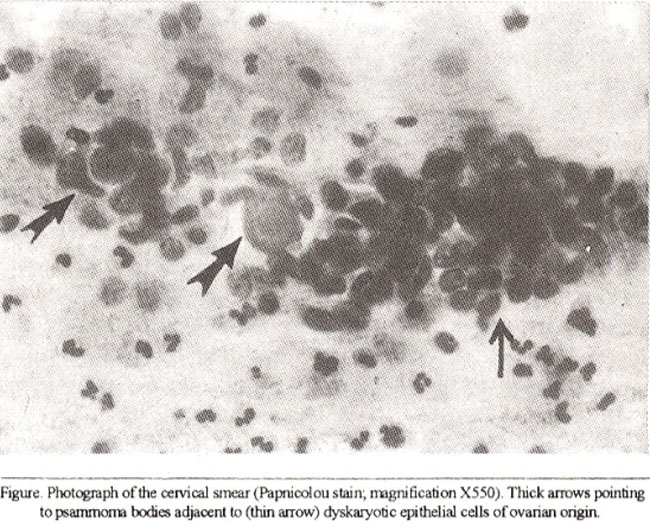
In the light of the discrepancy between the colposcopic findings and the repeat cytology, as well as the presence of psammoma bodies, it was decided to further investigate this woman by laparoscopy, hysteroscopy and cervical cone biopsy. At laparoscopy, both ovaries were enlarged to approximately 6 to 7 cm diameter and both had widespread cauliflower excrescences approximately 5 to 10 mm diameter. Similar seedling deposits were seen inthe pouch of Douglas (POD), on the uterosacral ligaments, on the fallopian tubes, on the back of the uterus and on the pelvic peritoneum A small amount of free fluid seen in the POD was aspirated and sent for cytological examination. Multiple biopsies of the cauliflower excrescences were taken. Hystmscopy was entirely normal. In the light of the laparoscopic findings, the cervical cone biopsy was deferred. The free fluid from the POD contained clumps of well differentiated glandular cells consistent with a serous cystadenonia. Histology of the tumour biopsies showed a papillary serous tumour, of borderline malignancy, containing numerous psammoma bodies. The woman and her husband were carefully counselled. Thebonlerline nature of the tumour was explained. The possibility of deferring definitive surgery to allow a pregnancy was also discussed. The couple volunteered that they had tried unsuccessfully to achieve a pregnancy over the preceding three years and had been on the verge of being referred for further investigation of their primary infertility. They felt that the time it might take to further investigate and any treatment givenfortheir infertility, mightjeopardise the chances of total cure of the ovarian lesion. They therefore opted to proceed to immediate surgical treatment and she underwent total abdominal hysterectomy, bilateral salpingo-oophorectomy and omentectomy. In addition to the findings at laparoscopy, numerous seedling deposits were seen on the abdominal parietal peritoneum, while the omentum, diaphragm and liverappeared to be free of tumour. She made an uncomplicated post-operative recovery. At histology, sections from the cervix showed non-specific inflammatory changes, with no evidence of CIN. Sections from both the ovaries showed the presence of bilateral papillary serous tumours of borderline malignancy associated with the presence of numerous psammoma bodies. Similar deposits were found in the omental and peritoneal sections. A diagnosis of bilateral ovarian papillary semus tumours of borderline malignancy with intraperitoneal spread was therefore made. She was referred to the Regional Consultant Oncologist, who decided to proceed to give her chemotherapy.
Discussion
The occurrence of psammoma bodies in Cervical Smears is rare. In a review of 234,318 smears, Kern1 found them in only seven. Three were associated with benign conditions, while the other four were associated with cancer. Although, classically associated with ovarian and thyroid carcinoma2,3 psammoma bodies are in fact found in a variety of neoplastic and non-neoplastic conditions. These include endometrial adenocarcinoma4-6, benign proliferative ovarian lesions and reparative processes involving the coelomic epithelium of ovaries and fallopian tubes7 and they may even be found in the meninges, choroid plexus, pineal body and thyroid gland in the absence of any pathology. They have also been reported in association with tuberculous endometritis8. intrauterine devices9,10 and in association with bening endometnal pathology in women on oral contraceptives11. Psanunoma bodies (from Greek psammos, meaning sand), are small round, concentrically laminated, often calcified structures. Their origin and development remains an enigma, although hypothesis include epithelial degeneration associated with cell death and changes in the pH of the milieu; mineralization of the secretions of the epithelial tumour cells and reflection of tumour-host reaction. The presence of psammoma bodies in cervical smear should be regarded as a stimulus for further investigation.
This case illustrates the management dilemmas which arise when malignancy occurs ma young nulliparous woman. It could be argued that laparoscopic biopsy was inappropriate ina situation where malignancy was suspected and that having taken that option, perhaps frozen section exanunation might have aided definitive treatment. Firstly, it was evident at laparoscopy that the tumour, whatever its nature had already spread throughout the pelvis and it was felt that biopsy was unlikely to spread it further. Secondly, it was decided that whatever the diagnosis given the woman’s age and nulliparity, it was prudent to obtain a definitive diagnosis and have the opportunity of counselling the woman, before proceeding to major surgery. The slight delay in treatment was unlikely to adversely affect the prognosis. With the benefit of hindsight, it is clear that frozen section would have raised difficulty in interpretation, since a definitive diagnosis of borderline malignancy requires examination of the whole specimen.
Borderline malignancy is a histological diagnosis made regardless of the clinical stage12. Prognosis is good, with 75% ten year survival having been recorded even in cases with residual disease13. Conservative management to allow child-bearing can therefore be considered. This option was discussed with the couple. However, they not unreasonably pointed to their failure to conceive overa three year period and were reluctant to embark on investigation and treatment where the time scale involved could not be determined. Furthermore, the effect on tumour progression of some current treatment, such as supra- physiological gonadotrophins given for ovulation induction are unknown. Information on borderline malignancies is sparse and the rate of progression of tumour in an individual patient cannot be determined. The opportunity to carefully and extensively counsel this woman and her husband, was welcomed by all concerned in her care and probably went along way to help her to come to terms with the radical surgery she had to undergo at such an early age. There was evidence of iritrapentoneal spread of this malignancy and it is evident that not all the tiny seedlings on the abdominal peritoneum were removed. Although this tumour progresses slowly, there are no reports of spontaneous regression. It is therefore, reasonabe to assume that in the long run these seedlings are likely to develop into more sinister lesions. It therefore seemed prudent to administer chemotherapy to this woman in an attempt to mop up residual seedlings and thereby perhaps effect total cure.
References
1. Kern, SB. Prevalence of psammoma bodies in Papanicolou-stained smears. ActaCytol., 1991;35:81-88.
2. Beyer-Boon, ME. Psammoma bodies in cervico-vaginal smears: An indicator of the presence of ovarian carcinoma. Acta Cytol., 1974,18:41-44.
3. Qizilbash, A.H. Ovarian carcinoma identified by psammoma bodies in vaginal smear. Can. Med. Assoc. J., 1974;1 10:185-186.
4. Fujimoto, I., Masubuchi, S., Miwa, H. eta!. Psammoma bodies in cervico-vaginal and/or endometrial smears. Acts Cytol., 1982;26:3 17-322.
5. Spjut, H. J., Kaufman, RH. and Carigg, S. S. Psammoma bodies in cervico-vagi. nal smears. Acts Cytol., 1977;21 :550-552.
6. Weaver, R.J. and Wilson, R. Endometrial carcinoma with psammoma bodies in thevaginal smear. Am. J. Obstet. Gynecol., 1967;97:869-870.
7. Picoff, R.C. and Meeker, C.I, Psammoma bodies in cervico.vaginal smear in association with benign papillary stmctures in the ovary. Acts Cytol., 1970; 14:45-47.
8. Jenkins, D.M. and Goulden, R. Psammoma bodies in cervical vagina! smears. ActaCytol., 1977;21:112-113.
9. Highman, W.J. Calcified bodies and the intrauterine contraceptive device. Acts Cytol., 1971;15:473-475.
10. Barter, J,F., Orr, J,W., Holloway, R.W. et al. Psammoma bodies in cervico-vagina! smear associated with intrauterine contraceptive device: A case report. J. Reprod. Med., 1987;32:147.148.
11. Valicenti, P. F and Priester, S. K. Psammoma bodies ofbenign endometrial origin in cervico-vaginal cytology. Acta Cyto!., 1977;21:550-552.
12. Ovarian tumour pane! ofthe Royal College of Obstetricians and Gynaecologists. Ovarian epithelial tumours of borderline malignancy: Pathological features and current status. Br. J. Obstet. Gynaecol., 1983;90:743-750.
13. Standing Committee on Cancer of The Standing Advisory Committee. Management of ovarian cancer; Current clinical practices. Report of a WorkingGroup, RCOG Publication, 1991.
Journal of the Pakistan Medical Association has agreed to receive and publish manuscripts in accordance with the principles of the following committees:


