

Malee S. Fernando ( Department of Pathology, Faculty of Medicine, University of Colombo, Colombo, Sri Lanka. )
MV Chandu de Silva ( Department of Pathology, Faculty of Medicine, University of Colombo, Colombo, Sri Lanka. )
June 1999, Volume 49, Issue 6
Case Reports

Introduction
Insular thyroid carcinoma (ITC) was first described as a distinct thyroid neoplasm by Carcangiu et al in 19841. This rare, tumour is aggressive and often lethal2. ITC is classified morphologically and biologically as a tumour intermediate between well differentiated (papillary and follicular) and undifferentiated (anaplastic) thyroid carcinomas2,3. There have been only a few publications on cytological findings in fine needle aspiration (FNA) cytology of ITC4-7. We report a case of ITC and discuss its fine needle findings.
Case Report
A 59-year female presented in 1997 with rapid enlargement of a long-standing nodular goitre. There was no history of exposure to radiation or family history of thyroid disease. Examination confirmed a multinodular goitre with hard nodules. There was no lymphadenopathy or distended neck veins. Thyroid function tests were normal and 1131 scan revealed cold nodules. FNA was done with a 23-gauge needle, fixed in 95% ethanol and stained with haematoxylin and eosin (H&E). The patient had .a total thyroidectomy and was referred to the oncologist for follow up. She remains well 22 months after surgery.
Pathological Findings
Thyroid FNA Cytology
The smears consisted mainly of large multilayered loosely cohesive nests of uniform cells (figure 1).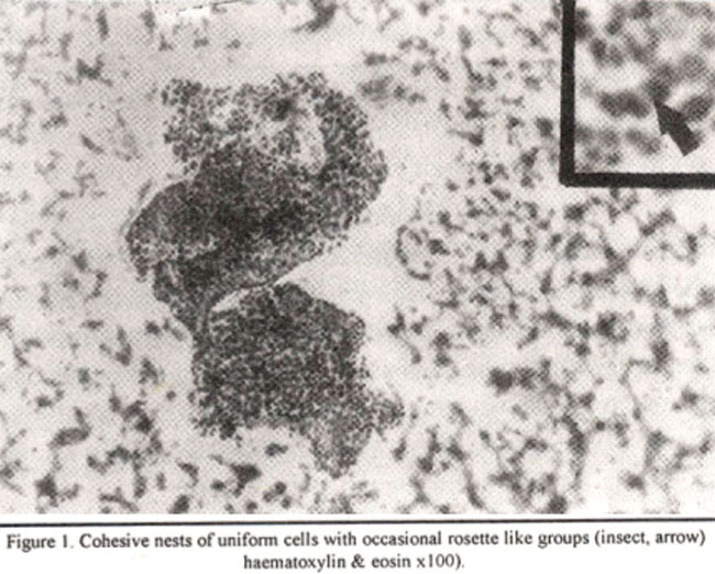
Most nests were solid with occasional rosette like groups (Figure 1 inset, arrow). There were no true microfollicles. The round to oval tumour cells were small (0.2-0.4 mm) With scanty amorphous cytoplasm. The nuclei were granular and hyperchromatic with small indistinct nucleoli. Nuclear grooves and orphan Annie type clearing were absent. Papillary features and giant cells were not identified
Thyroid Histology
The right lobe measuring 50x33x21 mm in maximum dimensions was replaced by a firm multilobated mass. Cut surface was almost replaced by two circumscribed, non-encapsulated, variegated firm tumour masses measuring 40 mm in maximum dimeter. On microscopic examination the tumour was composed of large round to ovoid, well defined nests of small uniform cells surrounded by a loose fibrovascular stroma (Figure 2).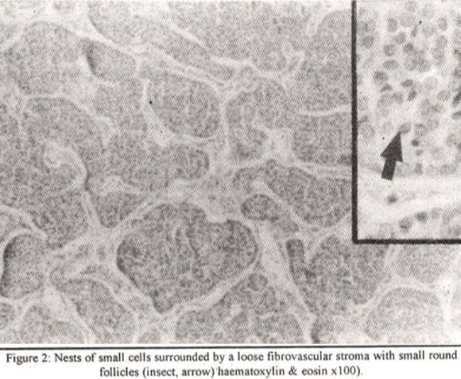
There was artifactual retraction of the stroma resulting in clefts beween tumour cells. Some nests contained small round haphazardly distributed follicles (figure 2 inset, arrow) giving a cribriform. pattern. The cells had hyperchromatic nuclei with indistinct nucleoli. Mitoses were sparse. Thyroid capsule was not penetrated. There was no necrosis, vascular emboli or features of a papillary neoplasm.
Discussion
Langhans in 1907 described a thyroid neoplasm composed of uniform cells arranged in a striking nesting pattern, with small cribriform lumina1. This tumour was recognised as a morphological entity with distinct biological behaviour in 19842. It has a clinical course in-between the often curable well differentiated papillary and follicular carcinomas and the highly lethal undifferentiated anaplastic carcinoma1-3. The disease is slightly more common in females3. The mean age at time of initial diagnosis is 55 years4. Metastases to regional nodes, lungs and bone has been reported and often resulted in death despite aggressive surgical, external beam therapy and chemotherapy8.
References
1. Carcangiu ML, Zampi G, Rosai J. Poorly differentiated (insular) thyroid carcinoma: A reinterpretation of Langhans’ “Wuchemde Struma”. American Journal of Surgical Pathology, l984;8:655-68.
2. Rosai J, Carcangiu ML, DeLellis RA. Atlas of tumour pathology. Tumours of the thyroid gland. Third series, fascicle 5. Washington DC. Armed Forces Institute of Pathology, 1 994;123-33,
3. Rosai J. Thyroid gland. hi: Ackerman’s surgical pathology. Volume 1. 8th edition. St. Louis. Mosby, 1996;532-534.
4. Paik SS, Kim WS, Hong EK, et al. Poorly differentiated (insular) carcinoma of the thyroid gland. Two case reports. J. Kr. Med. Assoc., 1997;12:70-74.
5. Zakowski MF, Schlesinger K, Mizrachi HH. Cytological features of poorly differentiated insular’ carcinoma of the thyroid. Acts Cytot. I992;36-523-6.
6. Sironi M, Collini P, Cantaboni A. Fine needle aspiration cytology of insular thyroid carcinoma. A report of four cases. Acta Cytol., 1992;36:435-39.
7. Kuhel WI, Kutler Dl, Santos-Buch CA. Poorly differentiated insular thyroid carcinoma. A case report with identification of intact insulae with needle aspiration biopsy. Acta Cytol., 1998;42:991-7.
8. Ashfaq R, Vuitch F, Delgado R, et al. Papillary and follicular thyroid carcinomas with an insular component. Cancer, 1994;73:416-23.
Journal of the Pakistan Medical Association has agreed to receive and publish manuscripts in accordance with the principles of the following committees:


