

Shahina Qureshi ( Children’s Hospital, Pakistan Institute of Medical Sciences, Islamabad. )
Asif Zia Khattak ( Children’s Hospital, Pakistan Institute of Medical Sciences, Islamabad. )
Naila Yaqub ( Children’s Hospital, Pakistan Institute of Medical Sciences, Islamabad. )
October 2000, Volume 50, Issue 10
Case Reports

Introduction
Congenital leukemia is a rare and poorly defined disease. Many authors define it as leukemia occurring from birth to four week1,2. Others recognize it as being synonymous with leukemia of the neonatal period and infancy3. It is commonly acute myeloblastic2,4 but may be acute lymphoblastic or juvenile myeloid in type5. It is associated with several trisomies, especially trisomy 213,6,7. Trisomy 13, trisomy 153 and several translocations such as translocation x:68, translocation 4:119 and monosomy 710.
Other associations include ElIis-Van-Creveld syndrome11, absence of radii12, patent ductus arteriosus, Klippel-fiel syndrome, atrial and ventricular septal defects12. Non-immune hydrops caused by congenital leukemia has also been reportede13.
One recognized peculiarity is the frequency of skin infiltration which is associated with poor prognosis14,15. Organ involvement is common and leads to hepatomegaly, splenomegaly and nephromegaly. Renal involvement can occur with or without dysfunction and is due to infiltration with leukemic cells or hypertrophy due to metabolic burden imposed by the malignancy. We are reporting an unusual case of congenital acute lymphoblastic leukemia presenting with bilateral renal masses.
Case Report
A live month old male infant was admitted to Children’s Hospital, Islamabad. on February 5, 1996 with history of diarrhea for three weeks, fever, difficulty in breathing and vomiting for four days. Abdominal distention was noticed three days before admission. Past history revealed recurrent chest infection, the first one at three and a half months of age which was treated with inject able Kananiycin. At four months of age he again developed cough and breathing difficulty, which was treated with injetable cefazolin. Blood transfusion was given for anemia twelve days before admission. He later developed diarrhea which was treated with metronidazole suspension and injectable ceftixone. As there was no improvement he was referred to Children’s Hospital.
He was a full term baby delivered by normal vaginal delivery. He was breast fed, supplementary foods were started at four months of age. Immunization was up to date. Development was normal. Family history was insignificant, three year old sister was healthy. There was no history of exposure to radiation or drugs in the mother.
General physical examination revealed a pale, irritable and sick looking infant weighing 6.9 kg and measuring 64 cms. Temperature was 11030F and blood pressure 100/70 mmHg. Systemic examination revealed a distended abdomen, liver palpable 6 ems below the right costal margin, spleen was not palpable. Both kidneys were palpable in the lumbar regions and were firm and non-tender. Chest, cardiovascular and central nervous systems were normal. There was no lymphadenopathy. There were no congenital malformations, petechiae or skin infiltrations.
Laboratory investigations showed a hemoglobin of 10.3 Gm/dl, white blood cell count was 5700/mm, No abnormal cells were seen in the peripheral smear. Blood urea nitrogen was 23 mg/d and creatinine 0.3 mg/dl. Platelet count was 13,000. Electrolytes blood sugar and arterial blood gases were normal.
Urine showed traces of albumin, numerous red blood cells and granular casts. Ultrasound abdomen showed bilaterally enlarged kidneys. Right kidney was 9.8 cms in length and left kidney 9.5 cms. IVP showed hydronephrosis on right side.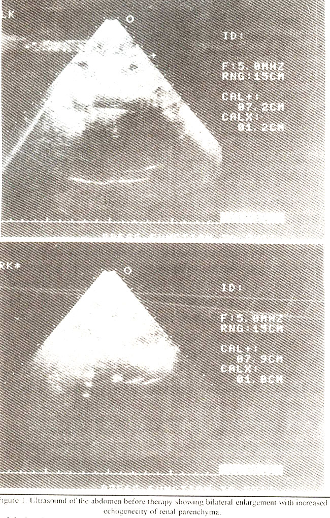
Urine and blood cultures were negative. Liver function tests were normal. Hepatitis B surface antigen were negative. During hospitalization the hemoglobin dropped and he developed thrombocytopenia. Bone marrow aspiration after one week showed acute lymphoblastic leukemia, LI type.
Treatment was started on February 19,1996 according to CCSG protocol 105 for high risk ALL with vincristine, L-asparginase, daunomycin and oral prednisolon. The child achieved complete remission in twenty eight days.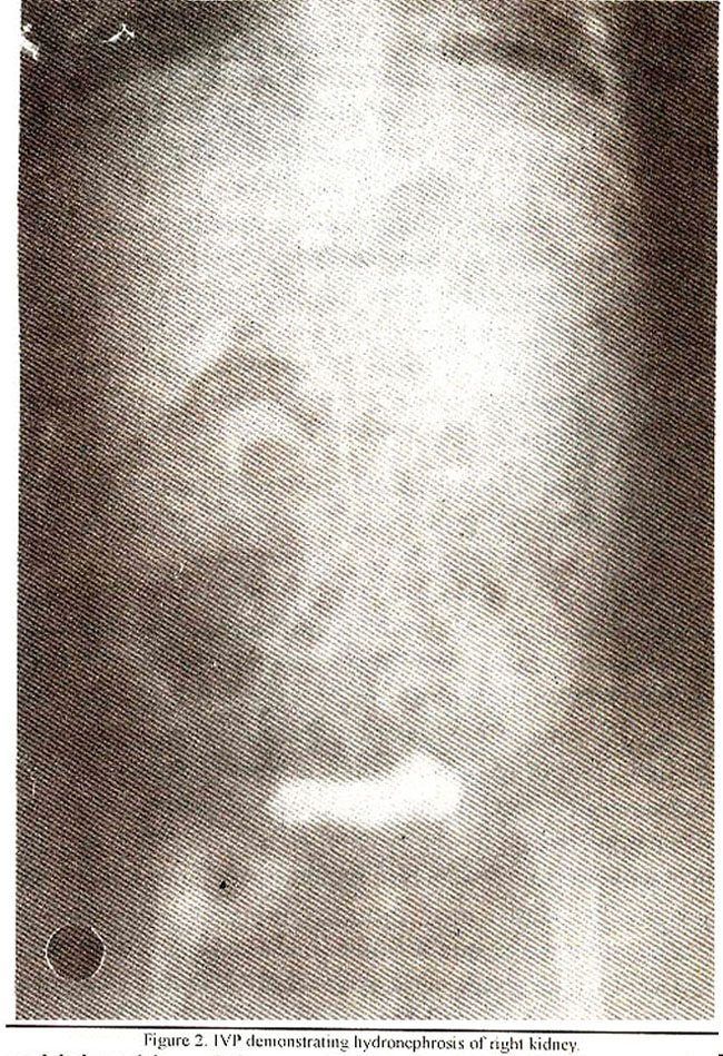
Abdominal ultrasound done fourteen days after treatment showed a reduction in size of both kidneys with right kidney measuring 7.9 cms and left kidney 7.2 ems, Consolidation therapy was given with cytosine arabinoside, four times a week for four weeks, intravenous cyclophosphamide on day I and 15, oral 6-mercaptopurine daily for twenty eight days, followed by interim maintenance with oral methotrexate once a week for four weeks, daily oral 6-mercaptopurine and weekly intrathecal methotrexate for four weeks. Intensification was done with vincristine once a week for three weeks, adriamycin once a week for three weeks, L-asparginase three times a week for two weeks and oral dexamethasone daily for twenty eight days. Maintenance therapy with oral 6-mercaptopurine daily, weekly oral methotrexate, vincristine and prednisolone every 28 days was started. He relapsed seven months after diagnosis and died due to pneumonia.
Discussion
Congenital leukemia and pseudoleukemia constitute a rare and poorly defined group of diseases3. The difficulty arises from the variation in criteria for recognition as congenital leukemia. Strictly speaking congenital leukemia should exist at birth. If diagnosed later, one should be reasonably certain that it was present at birth and would have been demonstrable if the appropriate diagnostic steps had been taken3.
There is a special and understandable tendency for clinicians to be more liberal in diagnosing congenital leukemia in patients with a family history of leukemia or associated congenital abnormalities. The best recognized of the latter is trisomy213,7,16 .
Leukemia that is strictly congenital is mostly acute myeloid, but may have a mixed picture with myeloid, lymphoid or erythroid progenitors17. Myeloid preponderance was seen in earlier reviews, where only four out of thirty-two patients were described as lymphocytic15 and two of twenty-one in another series were lymphocytic2. Excess of acute lymphoblastic leukemia has been recently reported in one series where, twenty-three out of thirty-two had acute lymphoblastic leukemia18. Juvenile chronic myelogenous leukemia is also relatively frequent during infancy19 and needs to be differentiated from a leukemoid reaction. Besides ALL and CML erythroleukemia has also been observed during this period17,20.
The clinical picture of leukemia in the neonate is different from that in older children4. Neonates usually present with purpura, hepatosplenomegaly and skin infiltrates in over half of the reported cases4. Many infants die due to respiratory distress secondary to pulmonary leukostasis and bronchopneumonia4. There is marked leukostasis and anaemia develops during the neonatal period. Platelets are usually reduced. The disease is not apparent at birth and signs may be evident after days or weeks. There may be an antecedent period of failure to thrive, diarrhea and low grade fever. Etiology of congenital leukemia is obscure4. Fetal X-ray exposure has not consistently been associated with an increased incidence of leukemia4.
Organ infiltration is common in leukemias and lymphomas21. Renal involvement is seen more frequently than in other non-renal malignancies, such as carinomas21. It is usually bilateral and confined to the cortex22. Renal function may be altered and is due to direct invasion of the kidneys, their vasculature or the collecting system by the tumours23. In 30% of the cases, renal enlargement is not due to malignant infiltration21. The cause is unknown, though tumor cells are seen on renal biopsy21. Rarely, a patient with renal failure of unknown origin and a normal peripheral blood count, has been found to have parenchymal infiltration due to ALL, on renal biopsy24. Despite the frequency of leukemic or lymphornatous infiltration of the kidney it may be recognized only on post mortem examination as reported in 60% of adults dying of leukemia25. The correlation of biopsy findings with the clinical picture is poor23. Azotemia, palpable flank mass, hypertension, albuminuria, hematuria and nephrotic syndrome may be present26. Excretory urography shows renal enlargement in 66% of cases, confirmed by tiltrasonography and computerized tomography26.
Renal involvement is also seen in congenital leukemia but there is a paucity of data on this subject. In one series, six cases were reported at autopsy over a period of seven years27. All cases were diagnosed as acute myeloid leukemia. Histological abnormalities were detected in the liver, lungs, spleen, pancreas, heart, intestine and kidneys. Although the response to chemotherapy in infants with congenital leukemia is poor28, spontaneous and permanent remissions may occur and are seen in infants with Downs syndrome4,6,16,21,29 They have also been reported with translocation 5:629. The course of the disease in infants who do not have Downs syndrome is most often short27, many patients dying within days or weeks of infection or haemmorhage2. Infants with congenital leukemia should be treated with chemotherapy like older children4. As renal invasion is also a. manifestation of disseminated neoplasia, chemotherapy is indicated. Radiotherapy to one kidney along with chemotherapy may help improve renal function24. Although prognosis is poor, with proper treatment complete remission can be achieved.
References
1. Jadhay MV, Deshmukh SD Aggarval PV. Congenital leukemia. Indian Pediatr I 988;25:101.
2. Pierce Ml. Leukemia in (lie newborn. Indian Pediatr 1959;54:691.
3. Henderson ES, Lister TA, Leukemia, 5th edition. Philadelphia. W.B. Saunders Co., 1990 p. 338.
4. Nailian DG, Oski, Congenital leukemia. Flematology of infancy and childhood. 3rd edition. Philadelphia, W.B. Saunders Co., 987 p. 708.
5. I lazani A, lIarak Y, Herant M, et. al. Congenital juvenile chronic myelogenous leukemia, therapeutic trial with interferon alpha-2. Med. Pediatr. Oncol.,1993.21:73.
6. Kurahashi H, tiara J, Yamura-Yagi K. et al. Transient abnormal inyclopoesis in Down’s syndronte. Leukemia-lymphoma. I 995;8:465-66.
7. Engel RR, Hammond D. Transient congenital leukeinut in seven infants with mongolism. J. Pediatr., 1964;66:303.
8. Camey LA, Kinney JS, Higgins RR, et al. x:6 translocation in a child wilt, congenital acute lyniphocytie leukemia Cancer, 992:69:700.
9. lleints S, l3ekassy AN. New structural chromosome arrangements in congenital leukem as. Leukemia, 1987; 1:116-7.
10. Shitara T, Suetake N, Yugaini 5, et at. A case of congenital leukemia with monosonly 7. Ann. I Hematot., I 996;65 :274-6.
11. Miller DR. Newstead GJ, Young LW. Perinatal leukemia with a possible variant of the Ellis-Van Creveld syndrome. J. Pediatr., 1969;74:300-1.
12. Stransky E. Perinatal leukemia. Aeta Pediatr.Acad. Sci. hung., 1967;8:121-2.
13. Donnenfeld AE Scott SC, Henselder H, ci at, Prenatally diagnosed non¬immune hydrops caused by congenital leukeinias. l’renat. Diag., 1994;14:721.
14. Moliná DN, Sanches Jl,, Lugo Somolinos A. The spectrum of cutanceus lesions in pediatric patients with teukemias. P.R. Health Sci, J., l994;13:24-6.
15. Reinman DR, Clemens RL and Pillsbury WA. Congenital acute leukemia, skin nodules, a first sign. J. Pediatr., 1995;46:415-6.
16. Rosner F, Lee SL. Down’s syndrome and acute letukeinias, mycloblastic or lymphoblastic. Am.J.Med,, 1972:53:203-4.
17. Heikinemo M, Pakkala S. Juvonen E, et at. Immuno arid cytochetnical charaetenzation of congenital leukemia; a ease report Med. Pediatr. Oneol.,1994;22:279-80.
18. Finkelstein JZ, Higgins OR, Rissmsman E, et al. Acute leukemia during the first year of life. Presentation, chemotherapy and clinical course. Clin, Pediatr.,1992;11:236-8.
19. Tan KH, Tan TS, Chronic myeloid leukenmias in children. Singapore Med.J 1968;9:39.
20. Lardi AA. Familial chronic myclocytic leukemia like syndrome probably of congenital origin. Acta Pediatr., 1994;83:558.
21. Frei E III, Fritz RD. Price E, et al, Renal and Imepatic enlargentent in acute leukemia. Cancer. 1963;16:1089-90.
22. Sterhy NH. studies in enlargememtt of leuketnie kidneys. Aesa Flematol.,1995:14:354.
23. Gilbert EF, Rice EC. Lechanx PA. Renal function in children with teukeuaia. Am.J.Dis.Child., 1956:93:150-2.
24. Barakat AY, Mnaytnneli LO. Acute lymphoblastie leukemia diagnosed by renal biopsy. J.tJrol., 1997;8:103-5.
25. Shapiro ill, Raunsay CC, Jacobsen I IG. et al. Renal involvement in leukemia and lymphoma in adults. Am.J. Rontgenol., 1962:88:928-30.
26. Richmond J, Shennan RS, Dianiond MD, et at. Renal lesions associated with malignant lymphomas Am.J.Med., 1 962;32:184-85.
27. Badhe P13, Sane SY. Congenital leukemia, Organ involvement in six autopsy eases, J. Postgrad. Med., 1992;38:127-9.
28. Embon CM, Goldstein KL, HaIl MR, et at. Congenital leukemia, comprehensive nursing care for a rare disease; a ease study. Neonatal Network, 1995; 14:7-8.
29. Mayer JL, Seashire MR. l-Iajjar FM. Translocation 5:6 associated will, spontaneosusly remmtttng congenital leukemia. Cancer Cenet. Cytogenet.,1995;81 :38-41.
Journal of the Pakistan Medical Association has agreed to receive and publish manuscripts in accordance with the principles of the following committees:


