

Mubasher Ikram ( Departments of Otolaryngology, The Aga Khan University, Karachi, Pakistan. )
Moghira Iqbal ( Departments of Otolaryngology, The Aga Khan University, Karachi, Pakistan. )
Muhammad Aslam Khan ( Departments of Medicine, The Aga Khan University, Karachi, Pakistan. )
Erum Khan ( Departments of Microbiology, The Aga Khan University, Karachi, Pakistan. )
Mahnaz Shah ( Departments of Ophthalmology, The Aga Khan University, Karachi, Pakistan. )
Raymond A. Smego Jr ( Departments of Medicine, The Aga Khan University, Karachi, Pakistan. )
May 2011, Volume 61, Issue 5
Original Article

Abstract
Objective: To study the disease spectrum and salient management features of 36 patients with histopathologically-confirmed rhinocerebral zygomycosis seen at our academic center over a 16-year period.
Methods: Retrospective review of patients admitted to the Aga Khan University Hospital in Karachi, Pakistan from January 1991 to December 2006 with histopathologically-confirmed zygomycosis of the head and neck.
Results: Mean patient age was 40 ± 5.0 years (range, 34-63 years), and 23 (64%) patients were male. Thirty-two (89%) patients were referred from clinical services other than otolaryngology. Underlying predisposing conditions included diabetes mellitus (21 patients), haematologic diseases (9), and renal failure (6). Twenty (55%) patients had limited sinonasal disease, ten (28%) had orbital involvement, and six (17%) had intracranial extension. All patients underwent rigid nasal endoscopy and biopsy, and black necrotic tissue was seen in 22 (61%) instances warranting endoscopic or open surgical debridement. Four of 6 patients undergoing open surgery required orbital exenteration. Overall patient survival was 56% (20/36 patients). Diabetic patients had improved survival (17/21, or 81%) compared to patients with haematologic disorders (3/9, or 33%) (p = 0.001). All six patients with intracerebral disease died. Eighteen of the 22 (82%) patients treated with surgery plus amphotericin B survived vs. two of 14 (14%) receiving amphotericin B alone (p < 0.001).
Conclusions: In rhinocerebral zygomycosis, an aggressive, multidisciplinary, diagnostic and therapeutic approach that utilizes CT or MRI staging, and combines endoscopic or open surgical debridement with amphotericin B-based antifungal therapy offers the best chance of recovery.
Keywords: Zygomycosis, Mucormycosis, Fungal infection, Rhinocerebral (JPMA 61:477; 2011).
Introduction
Zygomycosis (also known as mucormycosis and phycomycosis) is a fulminant and invasive opportunistic infection caused by fungi of the class Zygomycetes, order Mucorales, family Mucoracae, and genera Mucor, Absidia, Rhizomucor, Rhizopus, and Cunninghamella, and all agents have clinically indistinguishable presentations.1 The disease can present in a number of localized (pulmonary, cutaneous, or intestinal) or disseminated forms, although the most common and often lethal form is rhinocerebral zygomycosis, and it is associated with diabetes mellitus, metabolic acidosis, debilitation, and immunosuppressive diseases and/or treatments.2 In this article we herein review our 13-year experience with 36 cases of histopathologically-confirmed rhinocerebral zygomycosis at an academic center in Pakistan, and describe the salient management features of this life-threatening infection.
Patients and Methods
The medical records of patients, admitted to the Aga Khan University Hospital in Karachi, Pakistan from January 1991 to December 2006 with histopathologically-confirmed zygormycosis of the head and neck, were retrospectively reviewed. The diagnosis was made in all cases by the presence of non-septate branching hyphae on histopathologic sections. Clinical data extracted from inpatient medical records included presenting signs and symptoms, age, sex, diagnostic tests and sensitivities, treatment, and disease course and outcome. Rigid nasal endoscopy and biopsy was performed on all patients, and tissue was sent for histopathology, fungal smear (using potassium hydroxide), and culture on Sabouraud\\\'s dextrose agar with incubation at 25ºC and 37ºC. Computed tomography (CT) scanning of the paranansal sinuses and brain, including axial and coronal sections was performed in all cases to determine extent of the disease. An infectious diseases specialist was involved early in the management of all subjects, and in cases with eye involvement preoperative and intraoperative opinions from an ophthalmologist were obtained regarding the viability of the globe. Following surgery, all patients were followed closely for disease progression and repeat debridement was performed where necessary. Surgery was not performed on patients with intracranial extension. In patients with large defects following aggressive surgery, reconstruction was undertaken using loco-regional flaps to cover defects. Patients who underwent maxillectomy and/or orbital exenteration required some type of prosthesis to cover the anatomic defects.
Results
Thirty-six patients with rhinoorbitocerebral zygomycosis were seen at our academic center during the study period; clinical and treatment characteristics for all cases are shown in Table-1.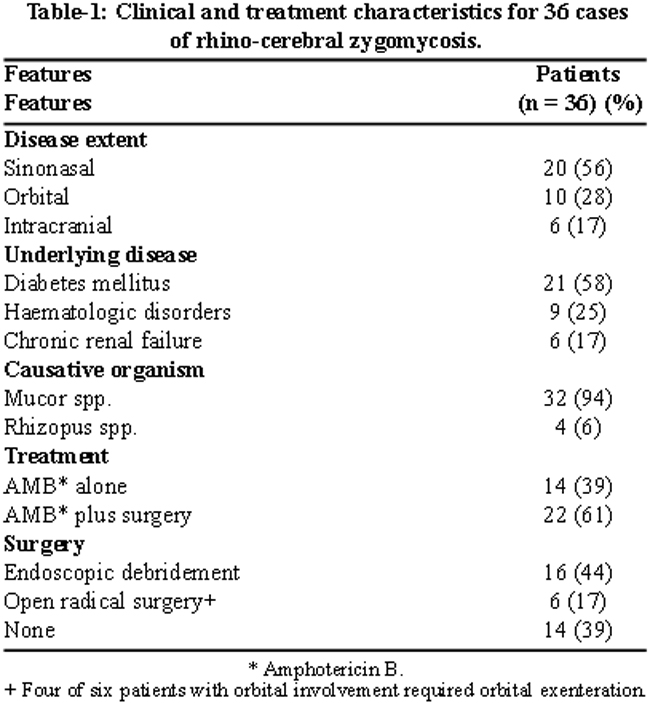
Mean patient age was 40 ± 5.0 years (range, 34-63 years) and 23 (64%) patients were male. Thirty-two (89%) patients were referred from clinical services other than otolaryngology including internal medicine, infectious diseases, endocrinology, ophthalmology, and oncology Predisposing conditions included diabetes mellitus (21 patients; 58%), haematologic diseases (9 patients; 25%), and renal failure (6 patients; 17%). All patients had rigid nasal endoscopy and biopsy which revealed black necrotic tissue in 22 (61%) instances, thereby warranting endoscopic or open surgical debridement. In 36 patients, a presumptive diagnosis of zygomycosis was made according to characteristic histopathology. Confirmatory tissue smears were positive for fungal hyphae in 22 (61%) cases, and 28 (78%) had operative tissue cultures positive for fungal growth. Causative microorganisms included either Mucor species (32 isolates) or Rhizopus species (4 isolates).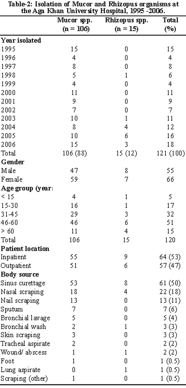
Table-2 shows isolation data for all zygomycetes at the Aga Khan University Hospital Mycology Laboratory during the period 1995-2006.
Twenty (56%) patients had disease limited to sinonasal regions, ten (28%) had evidence of involvement of the orbit at the time of initial presentation, and six (17%) patients had intracranial extension. All patients received amphotericin B within 48 hours of presentation based upon clinical suspicion of the disease. Most patients received a cumulative dose of 2-3 g of amphotericin B during the course of their treatment. A majority received maintenance iv amphotericin B infusions at home by home care nurses or in the hospital day care unit for periods up to several months depending on the clinical and radiologic response to therapy and degree of drug-related side effects. Almost all patients tolerated drug therapy well without any prolonged interruptions in therapy, and none suffered any significant long-term adverse drug effects. Nephrotoxic effects of amphotericin B were reversible and managed by ensuring adequate daily hydration with iv normal saline prior to daily infusions, and by withholding therapy for a few days during periods of transient escalating azotaemia.
Endoscopic debridement alone was performed in 16 (44%) patients, and six (17%) subjects required open/radical surgical procedures like Caldwell-Luc, facial debridement, and/or hemimaxillectomy. Four of the patients undergoing open surgery required orbital exenteration. Three patients required more then one surgical debridement, and one patient underwent three debridements. In fourteen patients (39%) surgery was not performed due to an advanced stage of disease or poor medical condition, or due to refusal by the patient or family.
Clinical outcomes for all 36 patients with invasive zygomycosis are summarized in Table-3.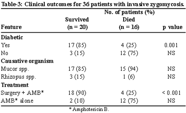
The overall patient survival was 56% (20 of 36 patients). Among the 16 deaths, six patients had intracranial extension of disease and six had underlying haematologic disorders (including one patient with extensive faciocutanous zygomycosis and acute leukaemia) and four had chronic renal failure. Diabetic patients had a better outcome in our series: 17 of 21 (81%) survived compared to 3 of 9 (33%) patients with underlying haematologic conditions (p = 0.001). All six patients with intracranial extension died. Eighteen of 22 (82%) patients treated with surgery plus amphotercin B survived vs. two of 14 (14%) receiving amphotercin B alone (p < 0.001).
Discussion
Zygomycosis involving the upper airway was first described in 1885 by Paltauf, who named it mycosis mucornia.3 In 1943, Gregory and colleagues reported the characteristic findings of advanced rhinocerebral zygomycosis in three patients with diabetic ketoacidosis4 and Harris reported the first cure of this disease in 1955.5
Timely diagnosis and successful management of mucormycosis requires a coordinated, multidisciplinary approach employing reversal of underlying immunosuppression, correction of metabolic acidosis, aggressive surgical intervention and systemic antifungal therapy, and the assistance of otolaryngologists, ophthalmologists, radiologists, internists, and infections disease and/or oncology specialists. Zygomycosis should be regarded as a medical-surgical emergency; antifungal therapy should be initiated at the slightest suggestion of infection, and patients should undergo endoscopic evaluation without delay, in order to visualize sinonasal structures and obtain appropriate specimens for histopathologic and microbiologic sampling. Direct nasal smear examination and fungal cultures are helpful in diagnosis, but if negative do not exclude the disease. Intraoperative frozen section is a specific and sensitive method of making a rapid diagnosis of rhinocerebral zygomycosis. In tissue biopsy, identification of non-septate, branching (at 90 degrees) hyphae without granuloma formation confirms the diagnosis. Fine-needle aspiration cytology appears to represent a useful alternative to tissue biopsy.6 Computed tomography or magnetic resonance imaging (MRI) is recommended for staging the extent of disease, and infiltration of periantral fat planes may represent the earliest imaging evidence of invasive fungal disease and should suggest the possibility of invasive fungal sinusitis in the appropriate clinical setting.7 Adequate debridement involves removal of all devitalized tissue up to healthy or bleeding margins,7 and multiple debridements may be necessary in some patients.1
Amphotericin B is the drug of choice for medical management of zygomycosis, although side effects including nephrotoxicity may require dose adjustment or limit use. Lipid-based formulations such as liposomal amphotericin B, amphotericin B lipid complex,8,9 and amphotericin B colloidal dispersion10,11 can be given in higher doses than amphotericin B deoxycholate and are associated with reduced side effects, most importantly dose-limiting renal toxicity. The imidazole, itraconazole, has in vitro activity against the etiologic agents of mucormycosis, but there is extremely limited clinical experience with its use.12 In vitro and in vivo resistance of zygomycetes to the triazole, voriconazole, have been associated with mucormycosis breakthrough in immunosuppressed populations in which the drug is used for antifungal prophylaxis.13 In the largest collection of in vitro susceptibility data for clinical isolates of zygomycetes, amphotericin B was the most active antifungal agent tested.14 The significant in vitro activity of the new triazole, posaconazole, against several species appears to support its reported, albeit limited, clinical efficacy. Early case reports indicate that posacanzole may be a useful drug for patients with rhinocerebral zygomycosis, including disease refractory to amphotermicn B.15-17 Other rarely reported adjuncts to systemic antifungal therapy for mucormycosis include nebulized amphotericin B, granulocyte macrophage-colony stimulating factor, and hyperbaric oxygen therapy.18-20
Good prognostic factors for rhinocerebral zygomycosis include early diagnosis and initiation of treatment21 and correction of underlying metabolic acidosis,22 while poor prognostic features include facial necrosis and nasal deformity21 and intracranial extension.21,23 Unlike an earlier report but similar to findings reported by Blitzer et al,21 we found that patients with diabetes mellitus had a better outcome than those with haematologic disorders including leukaemia.23 Early diagnostic markers of infection include perinasal cellulitis and paresthesia, periorbital oedema, mucopurulent rhinorrhea, and nasal crusting.7
Our successful results with endoscopic sinus surgery confirms those previously reported in the literature, and demonstrate that this modality can be used alone or in combination with traditional open surgical procedures.24 The 82% survival rate seen in our patients with rhinocerebral zygomycosis treated with surgery plus antifungal therapy was similar to results reported in a study of 20 patients from Taiwan.25 Fifteen of 16 (94%) patients reported by Khor and coworkers who received surgical debridement combined with amphotericin B survived, compared to only one (20%) of 5 patients who received amphotericin B alone (p = 0.004).25 A delay in diagnosis was also associated with higher disease mortality (p = 0.028). Only 3 of 12 (25%) patients with rhinocerebral disease from Greece survived their infection,2 and all six of our patients with intracranial extension died.
Conclusion
In conclusion, intracranial extension and underlying haematologic disorders are poor prognostic factors in patients with rhinocerbral zygomycosis. An aggressive, multidisciplinary, diagnostic and therapeutic approach that utilizes CT or MRI staging, and combines endoscopic or open surgical debridement with amphotericin B-based antifungal therapy offers the best chance of recovery.
Conflicts of Interest:
None of the authors has any conflict of interest, financial or otherwise.
References
1.Ferguson BJ. Mucormycosis of the nose and paranasal sinuses. Otolaryngol Clin North Am 2000; 33: 349-65.
2.Petrikkos G, Skiada A, Sambatakou H, Toskas A, Vaiopoulos G, Giannopoulou M, Katsilambros N. Mucormycosis: Ten-year experience at a tertiary care center in Greece. Eur J Clin Microbiol Infect Dis 2003; 22: 753-6.
3.Paltauf A. Mycosis mucorina. Virchows Arch 1885; 102: 543-64.
4.Gregory JE, Golden A, Haymaker W. Mucormycosis of the central nervous system. A report of three cases. Bull Johns Hopkins Hosp 1943; 73: 405-15.
5.Harris JS. Mucormycosis: Report of a case. Pediatrics 1955; 16: 857-67.
6.Deshpande AH, Munshi MM. Rhinocerebral mucormycosis diagnosis by aspiration cytology. Diagn Cytopathol 2000; 23: 97-100.
7.Dhiwakar M. Thakar A, Bahador S. Improving outcomes in rhinocerebral mucormycosis: Early diagnostic pointers and prognostic factors. J Laryngol Otol 2003; 117: 861-5.
8.Handzel O, Landau Z, Halperin D. Liposomal amphotericin B treatment for rhinocerebral mucormycosis: How much is enough? Rhinology 2003; 41: 184-6.
9.Saltoglu N, Tasova Y, Zorludemir S, Dundar IH. Rhinocerebral zygomycosis treated with liposomal amphotericin B and surgery. Mycoses 1998; 41: 45-9.
10.Strasser MD, Kennedy RJ, Adam RD. Rhinocerebral mucormycosis, Therapy with amphotericin B lipid complex. Arch Intern Med 1996; 156: 337-9.
11.Moses AE, Rahav G, Barenholz Y, Elidan J, Azaz B, Gillis S, et al. Rhinocerebral mucormycosis treated with amphotericin B colloidal dispersion in three patients. Clin Infect Dis 1998; 26: 1430-3.
12.Parthiban K, Gnanaguruvelan S, Janaki C, Sentamilselvi G, Boopalraj JM. Rhinocerebral zygomycosis. Mycoses 1998; 41: 51-3.
13.Marty FM, Cosimi LA, Baden LR. Breakthrough zygomycosis after voriconazole treatment in recipients of hematopoietic stem-cell transplants. N Engl J Med 2004; 350: 950-2.
14.Almyroudis NG, Sutton DA, Fothergill AW, Rinaldi MG, Kusne S. In vitro susceptibilities of 217 clinical isolates of zygomycetes to conventional and new antifungal agents. Antimicrob Agents Chemother 2007; 51: 2587-90.
15.Rutar T, Cockerham KP. Periorbital zygomycosis (mucormycosis) treated with posaconazole. Am J Ophthalmol 2006; 142: 187-8.
16.Kok J, Gilroy N, Halliday C, Lee OC, Novakovic D, Kevin P, Chen Sl. Early use of posaconazole in the successful treatment of rhino-orbital mucormycosis caused by Rhizopus oryzae. J Infect 2007; 55: e33-6.
17.Gelston CD, Durairaj VD, Simoes EA. Rhino-orbital mucormycosis causing cavernous sinus and internal carotid thrombosis treated with posaconazole. Arch Ophthalmol 2007; 125: 848-9.
18.Garcia-Diaz JB, Palau L, Pankey GA. Resolution of rhinocerebral zygomycosis associated with adjuvant administration of granulocyte-macrophage colony-stimulating factor. Clin Infect Dis 2001; 32: e145-50.
19.Kajs-Wyllie M. Hyperbaric oxygen therapy for rhinocerebral fungal infection. J Neurosci Nurs 1995; 27: 174-81.
20.Hamilton JF, Bartkowski HB, Rock JP. Management of CNS mucormycosis in the pediatric patient. Pediatr Neurosurg 2003; 38: 212-5.
21.Blitzer A, Lawson W, Meyers BR, Biller HF. Patient survival factors in paranasal sinus mucormycosis. Laryngoscope 1980; 90: 635-48.
22.Shpitzer T, Stern Y, Anavi Y, Segal K, Feinmesser R. Mucormycosis: Experience with 10 patients. Clin Otolaryngol Allied Sci 1995; 20: 374-9.
23.Butugan O, Sanchez TG, Goncalez F, Venosa AR, Miniti A. Rhinocerebral mucormycosis: Predisposing factors, diagnosis, therapy, complications and survival. Rev Laryngol Otol Rhinol (Bord) 1996; 117: 53-5.
24.Jiang RS, Hsu CY. Endoscopic sinus surgery for rhinocerebral mucormycosis. Am J Rhinol 1999; 13: 105-9.
25.Khor BS, Lee MH, Leu HS, Liu JW. Rhinocerebral mucormycosis in Taiwan. J Microbiol Immunol Infect 2003; 36: 266-9.
Related Articles
Journal of the Pakistan Medical Association has agreed to receive and publish manuscripts in accordance with the principles of the following committees:


