

Imaad-ur-Rehman ( Department of Diagnostic Radiology, Aga Khan University Hospital, Karachi. )
Wasim Memon ( Department of Diagnostic Radiology, Aga Khan University Hospital, Karachi. )
Yousuf Husen ( Department of Diagnostic Radiology, Aga Khan University Hospital, Karachi. )
Waseem Akhtar ( Department of Diagnostic Radiology, Aga Khan University Hospital, Karachi. )
Raafay Sophie ( Department of Diagnostic Radiology, Aga Khan University Hospital, Karachi. )
Muhammad Akbar Baig ( Department of Diagnostic Radiology, Aga Khan University Hospital, Karachi. )
January 2011, Volume 61, Issue 1
Original Article

Abstract
Objective: To evaluate accuracy of Computed Tomography (CT) for diagnosing malignancy in solitary pulmonary lesions (SPLs).
Methods: A prospective cross-sectional study was conducted from 20-01-2007 to 30-06-2008 at the Radiology department, Aga Khan University Hospital (AKUH) Karachi. Fifty-three patients with solitary pulmonary lesions (SPLs) seen in prior chest x-rays or chest CT scans were referred to radiology department for CT guided biopsy. CT scan was performed for each patient prior to biopsy and CT evaluation of the SPLs was performed followed by CT guided Biopsy. Histopathological diagnosis of the lesion was taken as the gold standard.
Results: CT was found to be 100% sensitive, 30% specific and 87% accurate for diagnosing malignancy in solitary pulmonary lesions while PPV and NPV were 86% and 100% respectively.
Conclusion: CT scan is highly sensitive yet non-specific and cannot be used as the definitive diagnostic modality for diagnosing malignancy in solitary pulmonary lesions.
Keywords: Computed Tomography, Solitary pulmonary lesion (JPMA 61:48; 2011).
Introduction
Cancer registries have documented lung cancer as the most common cancer in the world since 1985. By 2002 there were 1.35 million new cases, representing 12.4% of all new cancers. Globally it was also the most common cause of death from cancer, with 1.18 million reported deaths, or 17.6% of the world total.1 In Pakistan cancer of the lung has been ranked the most frequent malignancy in men in Karachi in the entire 1995-2002 period. In the 1998-2002 period the incidence rate increased to 25.5 per 100,000 (males) and 4.2 per 100,000 (females).2
The goal of radiologic evaluation of suspected pulmonary lesions, especially solitary nodules and masses, is to noninvasively differentiate between benign from malignant as accurately as possible. This is important because a solitary pulmonary lesion (SPL) is the initial radiographic finding in 20%-30% of patients with lung cancer, and prognosis depends in part on stage at presentation.3 Standard radiologic evaluation of a suspicious pulmonary lesion includes careful review of findings at chest radiography and, when appropriate, comparison with findings at prior radiography, chest fluoroscopy, CT and correlation with clinical signs and symptoms. When indicated and if available MRI, PET, and SPECT have also been utilized.4
Computed tomography (CT) is the imaging modality most often used to evaluate pulmonary nodules. The technique of helical low-dose CT scan is much more sensitive than a regular X-ray and can detect tumours if they are small.
Follow up by CT-guided percutaneous biopsy represents the most effective method to achieve a correct diagnosis in suspicious pulmonary lesions.5 It is widely accepted as effective and safe in many settings.6 It allows pathological verification of the radiological diagnosis with minimal invasiveness.7,8
Differential diagnosis of suspicious radiological pulmonary lesions also includes pulmonary metastases which are a common and frequent feature of a large variety of malignancies. Common causes of benign pulmonary lesions include among others, infectious granulomas most often due to Mycobacterium Tuberculosis and endemic fungi. According to the World Health Organization, in 2004 Pakistan pulmonary tuberculosis had a projected incidence of 181 per 100,000 population per year and a mortality rate of 41 per 100,000 population per year.9 The particularly variable presentations of pulmonary tuberculosis and its far greater prevalence in Pakistan can thus present serious pitfalls and errors in radiological evaluation.10
A need was therefore felt to determine the diagnostic accuracy of CT based diagnosis of malignancy in pulmonary lesions in our local setting.
Patients and Methods
Between January 2007 and June 2008 a prospective cross-sectional study was conducted. A total of 57 patients with solitary pulmonary lesions seen in prior chest x rays or chest CT scans were referred to radiology department AKUH for CT guided percutaneous biopsy. These patients included outside referrals as well as inpatients. Informed consent was obtained from all patients. Information regarding the patient\'s age, sex and pertinent clinical history were recorded.
All patients had 8-12 hours of fasting prior to the CT examination. Plain CT slices (10 mm interval) of the chest were obtained from lung apices to lung bases mainly focusing on the already known pulmonary lesion on single slice General electric scanner for every patient.
A team comprising of a radiologist with 3 years experience in chest CT and a senior radiology resident observed and documented the size of the lesion, location, nature of its margins, internal attenuation and presence/absence of cavitation, intranodular fat and intranodular calcium.
On the basis of these findings a CT diagnosis of the nature of the lesion i.e. either benign or malignant, was made using a set criteria.11,12
SPLs were scored as follows:11
a. Not suspicious for malignancy: homogenous, round, well-defined-margins, <3cm.
b. Low suspicion: Non homogenous, round, well-defined margins, <3cm.
c. Intermediate suspicion: non homogenous attenuation, well-defined margins, >3 cm.
d. Moderately high suspicion: non homogenous attenuation, irregular margins, > 3cm.
e. High suspicion: non homogenous attenuation, lobulated, spiculated margins, >3 cm.
SPLs in category a, b, c were considered benign and category d and e were considered malignant.
This was followed by CT guided percutaneous biopsy of the lesion performed in our department . Biopsy diagnosis obtained from the Pathology Department Aga Khan University Hospital, either benign/infection or malignant, was recorded.
Results
A total of 57 referred patients were found to have pulmonary lesions on CT scan in our department during the period of study, which were subsequently biopsied under CT guidance and had histopathology done in our hospital. Four of these biopsy specimens were diagnostically inadequate and a repeat biopsy was suggested which could not be performed for various reasons. Hence these 4 patients were omitted from the study. The data analysed is from the remaining 53 patients.
Of the 53 patients, there were 43 males and 10 females. A wide range of age was seen varying between 20 years and 83 years with mean age of 62 ± 13 years. For females the ages ranged between 50 and 74, with mean age of 62 ± 9 years, while in males the range was 20 - 83 years, with mean age of 62 ± 14 years.
Out of the 53 cases, 50 (94%) were read on CT as having positive likelihood for malignancy. On later histopathological examination 43 of the total 53 cases had pulmonary malignancies (81%) while 10 were diagnosed with benign pathologies. Among the male patients the malignancy rate was 84%, and in female cases it was 70%. The malignancies included non-small cell lung carcinoma (NSCLC, n = 30) (Figure-1),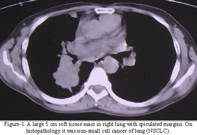
small cell lung carcinoma (SCLC, n = 4), lymphoblastic lymphoma (n =1), primitive neuroectodermal tumours (PNET, n =2), and six metastatic lesions including five from adenocarcinomas and one from renal cell carcinoma.
The histologically benign cases included tuberculous lesions (n =5) and one each of chondroid hamartoma, myxoid neoplasm, chronic inflammation, granular cell tumour and fibrous tissue. CT based diagnosis of benign lesion was made in 3 cases, all 3 of which proved benign on biopsy.
Thus there were 43 true positive, 7 false positive, 3 true negative and no false negative results reported on CT based assessment of likelihood of malignancy in these pulmonary lesions.
Statistical parameters thus showed sensitivity of 100%, specificity of 30%, positive predictive value of 86%, and negative predictive value of 100% and diagnostic accuracy of 87% for CT scan for diagnosing malignancy in solitary pulmonary lesions.
Irregular margins with spiculations and nonhomogenous attenuation were the most frequently seen findings in the positive cases (43 and 42 cases respectively). However irregular margins were also noted in 7 benign lesions and nonhomogenous attenuation noted in 6 benign lesions (Figure-2).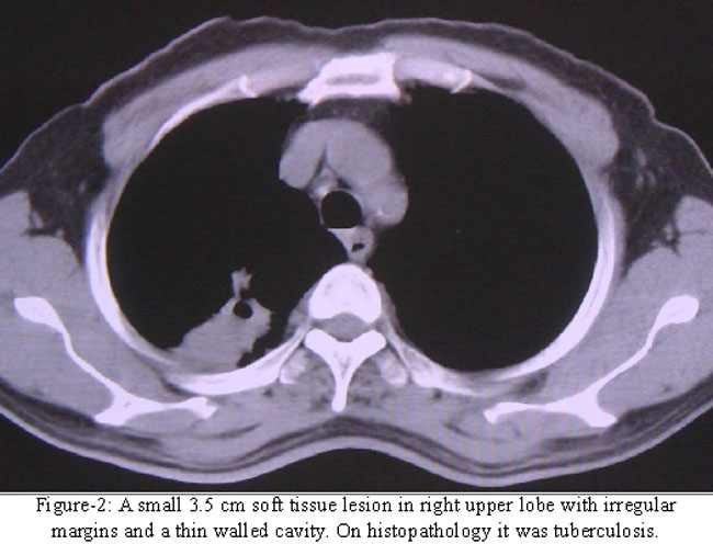
Size greater than 3 cm was noted in a total of 43 cases, 36 of which were malignant. Malignancy was also diagnosed in 7 of the 10 lesions less than 3 cm in size.
Discussion
In Pakistan at present, CT is the most commonly used non invasive modality for evaluating pulmonary lesions. It is therefore crucial to evaluate and verify the accuracy of CT in diagnosing malignancy so that we can plan the diagnostic workup of such patients in a realistic manner.
Our study demonstrates that CT scan based diagnosis is highly sensitive yet non-specific. All three major criteria to diagnose malignancy i.e. size of the lesion, border characteristics and internal attenuation are sensitive, but can be seen in a variety of benign conditions as well.
In our series, 43 of the 53 pulmonary lesions were greater than 3 cm in size and out of these 36 were found to be malignant (86%) while 10 lesions were less than 3 cm and out of these 7 lesions proved to be malignant (70%). This is in general agreement with published literature. In a study conducted by Steele et al,13 80 percent of solitary nodules larger than 3 cm in diameter were found to be malignant, compared to 20 percent of nodules less than 2 cm in diameter, while studies done by Zerhouni et al. and Siegelman et al. demonstrated that more than 90 percent of lesions larger than 3 cm are malignant.14,15
Regarding border characteristics in our study all 43 malignant lesions had irregular margins, a sensitivity of 100 percent; however, irregular margins were also seen in 7 of the 10 benign lesions. Hence this finding is only 30% specific. Overall accuracy of this finding was calculated to be 87 %. Conversely the sign of smooth margins was highly specific for benign nature of the lesion in our cases as only 3 lesions were seen to have smooth borders and all 3 were benign. However in this case the sensitivity was low as 7 of the 10 benign lesions did not show smooth margins. Rigler et al showed that a smooth border has a 20 percent likelihood of representing a malignant nodule. The likelihood increases to 60 percent with a scalloped border, 90 percent with spiculations and 95 percent in the presence of corona radiate.16
The third major characteristic of internal attenuation also had high sensitivity for malignant lesions yet was seen in a variety of benign lesions as well.
The most common pathology mimicking malignancy was tuberculosis. All five cases of tuberculosis in our study were read as malignant based upon CT findings. This depicts the extremely varied appearance of tuberculous lesions making it virtually unidentifiable based upon CT characteristics (Figure-2). Cherian et al in his study of atypical pulmonary patterns of tuberculosis found that the most common of these unusual radiographic patterns of tuberculosis was mass-like densities, most of which were initially and mistakenly diagnosed as neoplasms.17 Similar was the result in our study and is a major concern in our set up since TB has high prevalence in Pakistan.9
Comparatively low specificity seen in our study as compared to other studies18,12 is primarily attributable to these tuberculous lesions. We failed to identify any criteria which could improve the sensitivity or specificity for recognizing atypical TB lesions on CT scan. However only five cases of tuberculosis were seen in a total of fifty three patients which is too small a sample size to draw any realistic conclusion. This is a limitation in our study.
Another limitation was that no dynamic imaging was performed after contrast administration. The study was conducted in a single centre and only those patients were included who had known SPLs on prior radiographic evaluation thus creating selection bias in the study. No long term follow-up of patients is available.
Conclusion
Due to its low specificity, CT scan cannot be used as the definitive diagnostic modality for diagnosing malignancy in solitary pulmonary lesions, however it can be used as a localizing tool prior to CT guided percutaneous biopsy which is highly specific and sensitive.
References
1.Parkin DM, Bray F, Ferlay J, Pisani P. Global cancer statistics, 2002. CA Cancer J Clin 2005; 55: 74-108.
2.Bhurgi Y, Bhurgi A, Usman A, Sheikh N, Faridi N, Malik J, et al. Patho-epidemiology of lung cancer in Karachi (1995-2002). Asian Pac J Cancer Prev. 2006; 7: 60-4.
3.Viggiano RW, Swensen SJ, Rosenow EC 3rd. Evaluation and management of solitary and multiple pulmonary nodules. Clin Chest Med 1992; 13: 83-95.
4.Hartman TE. Radiologic evaluation of the solitary pulmonary nodule. Radiol Clin North Am 2005; 43: 459-65
5.Chojniak R, Isberner RK, Viana LM, Yu LS, Aita AA, Soares FA. Computed tomography guided needle biopsy: experience from 1,300 procedures. Sao Paulo Med J 2006; 124: 10-4.
6.Rossi ED, Mule A, Maggiore C, Miraglia A, Lauriola L, Vecchio FM, et al.. Cytologic diagnosis of pulmonary lesions. Rays 2004; 29: 357-61.
7.Mehmood N, Rizvi N, Khan RA, Shah RH. Role of Computed Tomography-Guided Percutaneous Transthoracic Needle Lung Biopsy in the Diagnosis of Peripheral Pulmonary Mass Lesion. J Coll Physicians Surg Pak 2001; 11: 482-5.
8.Qureshi SS, Bukhari MH, Hanif G, Azhar M, Iqbal N, Naveed IA. Accuracy and clinical utility of Cytological Typing of Lung Tumours. Ann King Edward Med Coll 2002; 8: 204-6.
9.World Health Organisation: Global Tuberculosis Control. WHO Report. 2006: Geneva: World Health Organisation, 2006.
10.Khan MB, Mashhood AA, Qureshi WA, Ibrar K. Tuberculosis - Disease pattern & the sputum microscopy yield. Pak J Chest Med Dec 2005; 11: 11-9.
11.Dahnert W. Radiology Review Manual. 6th ed. Philadelphia: Lippincott Williams and Wilkins, 2007; pp 424-9.
12.Kim SK, Allen Auerbach M, Goldin J, Fueger BJ, Dahlbom M, Brown M et al. Accuracy of PET/CT in characterization of solitary pulmonary lesions. J Nucl Med 2007; 48: 214-20.
13.Steele JD. The solitary pulmonary nodule. Report of a cooperative study of resected asymptomatic solitary pulmonary nodules in males. J Thorac Cardiovasc Surg 1963; 46: 21-39.
14.Zerhouni EA, Stitik FP, Siegelman SS, Naidch DP, Sagel SS, Proto AV, et al. CT of the pulmonary nodule: a cooperative study. Radiology 1986; 160: 319-27
15.Siegelman SS, Khouri NF, Leo FP, Fisherman EK, Braverman RM, Zerhouni EA. Solitary pulmonary nodules: CT assessment. Radiology 1986; 160: 307-12
16.Rigler LG. An overview of cancer of the lung. Semin Roentgenol 1977; 12: 161-4.
17.Cherian MJ, Dahniya MH, Al-Marzouk NF, Abel A, Bader S, Buerki K, Mahdi OZ. Pulmonary tuberculosis presenting as mass lesions and simulating neoplasms in adults. J Med Imaging Radiation Oncol 1998; 42: 303-8.
18.Lu G, Wang Z, Zhu H, Chang L, Chen Y, Wu J, et al. The Advantage of PET and CT Integration in Examination of Lung Tumours. Int J Biomed Imaging 2007; 2007: 17131.
Related Articles
Journal of the Pakistan Medical Association has agreed to receive and publish manuscripts in accordance with the principles of the following committees:


