

S. Sharieff ( Department of Adult Cardiology, National Institute of Cardiovascular Diseases, Karachi. )
K. S. Zaman ( Department of Adult Cardiology, National Institute of Cardiovascular Diseases, Karachi. )
July 2003, Volume 53, Issue 7
Original Article

Introduction
Peripartum cardiomyopathy (PPCM) is a rare form of left ventricular failure that is poorly characterized and of unknown etiology. Ritchie and Virchow1,2 first described an association between puerperium and cardiac failure in the middle and late 1800s. Later in 1971 Demarkis and associates3 described it as the development of cardiac failure during the last month of pregnancy or 5 months after delivery in a patient with no demonstrable heart disease before the last month of pregnancy and no discernible etiology for heart failure. More recently, it has been suggested that impairment of left ventricular function and increased left ventricular end-diastolic dimension should be added as diagnostic criteria for PPCM.4,5 Despite of current management, the mortality rate remains high between 25-50%3,6 and nearly half of patients surviving the initial episode of cardiac failure continue to have persistent left ventricular dysfunction.6 We attempted to identify the prognostic factors of peripartum cardiomyopathy (PP-CMP) in Pakistani patients.
Patients & Methods
From 1st December 1999 to 30th August 2001, 35 consecutive patients diagnosed to have peripartum cardiomyopathy (PP-CMP) at the National Institute of Cardiovascular Diseases (N.I.C.V.D.) Karachi, Pakistan, were enrolled. In each case the clinical history was verified and the two-dimensional Doppler echocardiogram was done at the time of diagnosis and later repeated at the end of six months of treatment to evaluate the left ventricular function so as to assess the recovery or progress of the disease.
Patiens with heart failure within the last month of pregnancy or 5 months postpartum, absence of pre-existing heart disease, no determinable etiology, and Echocardiographic evidence of left ventricular dysfunction.3,5 were included in this study.
Exclusion criteria were pre-existing heart disease e.g. congenital, valvular, or ischemic heart disease, heart failure secondary to anemia, infections, metabolic and toxic causes, presence of any other systemic illness before pregnancy e.g. autoimmune disease, and any complication arising during delivery e.g. puerperal infection, septicemia, ante-partum/post-partum hemorrhage, pulmonary embolism, amniotic fluid embolism, etc which may lead to heart failure.
Standard treatment for congestive cardiac failure including dispirin, angiotensin converting enzyme inhibitor (ACE-I), diuretics (Frusemide and aldactone) and digoxin, was given to the patients and their responses were analyzed.
The follow up visits were carried out for at least 6 months for every individual patients as nearly half of deaths usually occur during the first 3 months 3,7, while those who recover usually do so during first 6 months from the time of diagnosis3 and those with persistent left ventricular dysfunction after 6 months usually have poor prognosis.3,7
Patients were divided into two groups depending upon their clinical status at follow-up. Group A consisted of patients who improved or recovered, and Group B consisted of patients who either died or deteriorated and remained in functional class 111 or 1V of NYHA.8,9 Echocardiogram was repeated at the end of six months to evaluate the left ventricular function and to assess the recovery or progress of the disease.
Statistical analysis was done in SPSS system. Data was analyzed in two-tailed Chi-square test and two-tailed fisher exact test. Student t-test and Pearson's correlation coefficient were used for comparison and correlation. The data in the text are expressed as the mean+standard deviation. P-value less than 0.05 was considered significant.
Results
The mean age at the time of diagnosis was 30.8 ± 6.74 years (range 17 to 40 years), with 68.6% above 30 years. Majority of the patients were multiparous (77.14%) and presented during the postpartum period (71.4%). PPCM was more commonly found in patients belonging to lower socio-economic class with poor nutritional status (77%), 25.7% of patients had recurrent PPCM while 8.6% were diabetics, 71.43% had normal babies while 28.57 patients had either dead baby at delivery or their babies expired soon after delivery (Table 1).
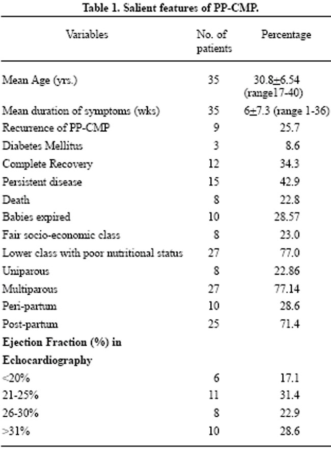
The mean duration of symptoms was 6 ± 7.3 (range 1-36) weeks at the time of diagnosis. Dyspnea was present in all patients followed by chest pain (45.71%), edema (37.14%) and paroxysmal nocturnal dyspnea (17.14%). The examination showed bilateral crepitations in the chest (88.7%), S3 gallop (65.71%), tachycardia (62.86%), edema (60%), hypotension (37.14%), raised JVP (28.57%) and anemia (2.86%). There was cardiomegaly on chest X-ray in 71.4%, while on ECGs 85.7% had poor 'R' wave progression in anterior chest leads and 5.7% had LBBB. Poor left ventricular function with ejection fraction <30% was found in 71.4% and 74% had dilated left ventricle with left ventricular end-diastolic dimension >55 mm; 34.3% of patients recovered completely while rest either had persistent and progressive disease (42.9%) or had expired (22.8%) (Figure).
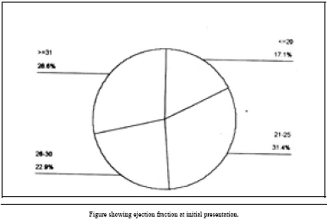
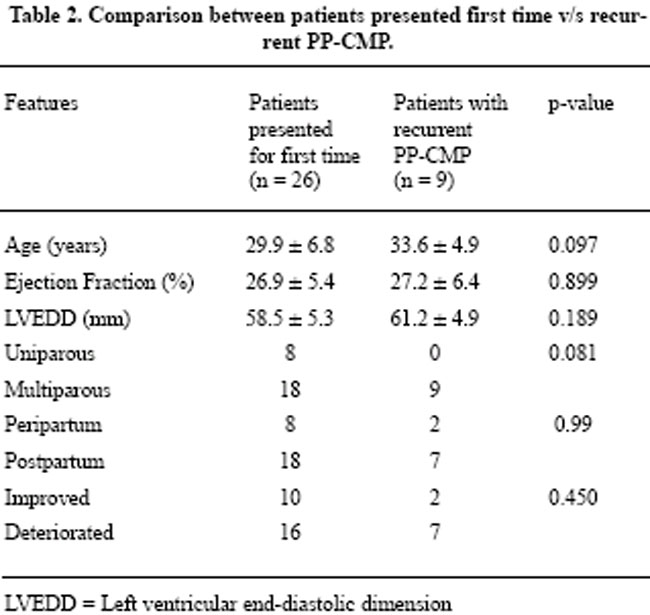
Table 2 shows that there was no significant difference (p values = 0.097, 0.899 and 0.189 respectively) between patients who presented for the first time v/s patients with recurrent PPCMP in the mean age, ejection fraction (EF) and left ventricular end diastolic dimension (LVEDD). Similarly, parity, time of onset (peri-/postpartum period) and outcome between these two groups were also statistically insignificant. To compare the various prognostic factors in PPCMP patients were divided in two groups depending upon their clinical status at the end of six months of follow-up. Group A consisted of 12 (34.3%) patients who improved and group B, 23 (65.7%) patients who deteriorated i.e., either expired or remained with persistent disease in functional class III or IV (Table 3).
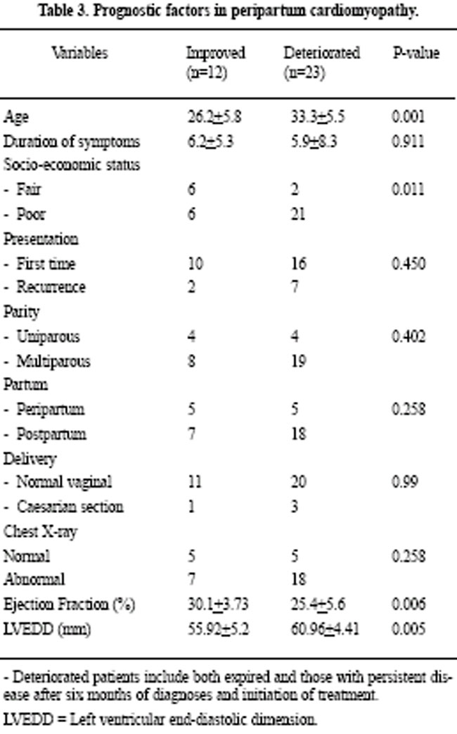
The patients who improved on therapy, as compared to those who deteriorated, were significantly younger (p-value <0.001), financially better off with better nutritional status (p value <0.011), had lower LVEDD (p value <0.005)and a better ejection fraction (p-value <0.006), while duration of symptoms, presentation period (first time or recurrent), parity, time of onset (peri-/post-partum), mode of delivery (caesarian section or normal vaginal delivery), chest X-ray and ECG abnormalities did not show significant relationship with outcome.
Deteriorated patients include both expired and those with persistent disease after six months of diagnoses and initiation of treatment. LVEDD = Left ventricular end-diastolic dimension.
Discussion
Peripartum cardiomyopathy is a rare cause of heart failure with high morality.3,6,7 The underlying cause remains undetermined, and diagnosis is essentially by exclusion; clinicians assuming that other problems such as volume overload, hypertension with systolic dysfunction, and sepsis, all of which occur at or after term, are absent. Evaluation by echocardiographic measurements of left ventricular chamber dimension and function at diagnosis and recovery; and by echocardiographic stress tests using pharmacologic stimulation with dobutamine to determine contractile reserve should be considered and recommended.6
The mean age in this study was 30.8 ± 6.54 years (range17-40) as most of our patients were >30 years of age (68.6%), emphasizing the fact that the disease has been found more commonly in women >30 years, although a wide range of ages have been reported.3,10 Also majority of our patients were multiparous (77.14%) which is consistent with some11 but different from other studies where they found the disease more commonly in younger age group with lower level of parity.8,12 Majority of our patients were from lower socio-economic class with poor nutritional status (77%), which is also one of the recognized risk factor for PPCM.11
Thus, the study showed advanced age (>30 years), lower socio-economic class with poor nutritional status, multiparity, past history of PPCM as the risk factors for peripartum cardiomyopathy, which patients are more prone to develop during postpartum period; these findings are same as reported in earlier studies.1, 3, 7,10,13,14 We also found that recurrent PPCM is more in advance age, as all of our 9 patients with recurrent PPCMP were above 30 years of age; among whom 2 (22.22%) recovered, 2 (22.22%) expired and 5 (55.56%) remained with persistent and progressive disease. Thus 77.78% of patients with recurrent PPCM either deteriorated or had expired by the end of six months of follow-up, showing that recurrent peripartum cardiomyopathy has much higher mortality and morbidity. Although there was no statistical significance between the mean age, ejection fraction (EF) and left ventricular end diastolic dimension (LVEDD), time of onset (peri-/post-partum), parity and outcome of patients with recurrent peripartum cardiomyopathy in our study as compared to those patients presenting for the first time with peripartum cardiomyopathy; however, these finding are consistent with the fact that patients, even after complete recovery from first episode of peripartum cardiomyopathy, may be at a higher risk for recurrence during subsequent pregnancies3,10,14,15 and the reason of this recurrence is still unknown. This is in contrast to Sutton et al16 that after resolution of left ventricular function the patients with PPCM are at no increased risk for cardiac problems on their subsequent pregnancies. Therefore, in our opinion the women with peripartum cardiomyopathy should be counseled for future pregnancies and be informed about the possibilities of recurrence of cardiomyopathy with subsequent pregnancies, which carries high mortality. However, if the pregnancy is still desired then patients should first undergo stress testing for example stress echocardiogram6,17 to evaluate for contractile reserve under hemodynamic stressful conditions.
The study showed advance age (>30 years), low socio-economic status with poor nutrition, lower ejection fraction (EF) and increased left ventricular end-diastolic dimension (LVEDD) as poor prognostic factors. However, Ravikishore8 did not find EF statistically significant in relation to prognosis, while Rosa et al15 did showed prognostic value in their study; however, they found parity, symptoms onset, cardio-thoracic ratio on chest X-ray (CXR) and hemodynamic data as significant markers of prognosis as opposed to our findings in which symptoms duration, presentation, parity, time of onset (peri/post-partum), mode of delivery and CXR were not statistically significant. Thus, in our study, those patients who did not improve on therapy were found to have higher left ventricular end-diastolic dimension with poor left ventricular ejection fraction on echocardiogram and advanced maternal age. It is also known that patients with persistent left ventricular dysfunction have an unacceptably high risk of cardiac complications and death during subsequent pregnancies and, therefore, should be counseled not to become pregnant again.18
In conclusion, we tried to describe the poor prognostic factors at initial presentation and to identify the high risk subjects and thus to provide effective treatment that will ensure a favorable return of normal left ventricular function.
References
1. Heider AL, Kuller JA, Strauss RA, Wells SR. Peripartum Cardiomyopathy: a review of the literature. Obstet Gynecol Surv 1999; 54:526-31.
2. Gouley BA, McMillan TM, Bellet S. Idiopathic myocardial degeneration associated with pregnancy and especially the puerperium. Am J Med Sci 1937; 19:185-99.
3. Demakis JG, Rahimtoola SH, Sutton GC, et al. Natural course of peripartum cardiomyopathy. Circulation 1971; 44:1053-61.
4. Manolio TA, Baughman KL, Rodeheffer R, et al. Prevalence and etiology of idiopathic dilated cardiomyopathy (summary of a National Heart, Lung and Blood Institute workshop). Am J Cardiol 1992;69:1458-66.
5. Hibbard JU, Lindheimer M, Lang RM. A modified definition for peripartum cardiomyopathy and prognosis based on echocardiography. Obstet Gynecol 1999; 94:311-16.
6. Lampert MB, Weinert BS, Hibbard J, et al. Contractile reserve in patients with peripartum cardiomyopathy and recovered left ventricular function. Am J Obstet Gynecol 1997;176:189-95.
7. Lampert MB, Lange RM. Peripartum cardiomyopathy. Am Heart J 1995;130: 860-70.
8. Ravikishore AG, Kaul UA, Sethi KK, et al. Peripartum cardiomyopathy: prognostic variables at initial evaluation. Int J Cardiol 1991;32:377-80.
9. Carvalho A, Brandao A, Martinez EE, et al. Prognosis in peripartum cardiomyopathy. Am J Cardiol 1989;64:540-42.
10. Seftel H, Susser M. Maternity and myocardial failure in African women. Br Heart J 1961;23:43-52.
11. Veille J. Peripartum cardiomyopathies: a review. Am J Obstet Gynecol 1984;148:805-18.
12. Ford RF, Barton JR, O'Brien JM, et al. Demographics, management and outcome of peripartum cardiomyopathy in a community hospital. Am J Obstet Gynecol 2000;182:1036-38.
13. O'Connell JB, Costanzo-Nordin MR, Subramanian R, et al. Peripartum cardiomyopathy: clinical, hemodynamic, histologic and prognostic characteristics. J Am Coll Cardiol 1986;8:52-6.
14. Witlin AG, Mabie WC, Sibai BM. Peripartum cardiomyopathy: an ominous diagnosis. Am J Obstet Gynecol 1997;176:182-8.
15. Rosa M, O'Connel JB. Peripartum cardiomyopathy in the 1980's: etiologic and prognostic considerations and review of the literature. Progr Cardiol 1989;212: 225-40.
16. Sutton MS, Cole P, Plappert M, et al. Effects of subsequent pregnancy on left ventricular function in peripartum cardiomyopathy. Am Heart J 1991;121:1776-8.
17. Lampert MB, Weinert BS, Hibbard J, et al. Contractile reserve in patients with peripartum cardiomyopathy and recovered left ventricular function. J Am Coll Cardiol 1994;23:428A.
18. Elkayam U, Tummala PP, Rao K, et.al. Maternal and fetal outcomes of subsequent pregnancies in women with peripartum cardiomyopathy. N Eng J Med 2001;344:1567-71.
Abstract
Objectives:
To describe the prognostic factors at initial presentation of patients with peripartum cardiomyopathy (PPCM).
Setting:
Department of Adult Cardiology, National Institute of Cardio-vascular Diseases Karachi, Pakistan, from December 1999 to August 2001.
Method:
A total of 35 patients diagnosed to have peripartum cardiomyopathy (PPCM) were studied. A detail clinical history was taken in each case and two-dimensional Doppler echocardiogram was done at the time of diagnosis and later repeated at the end of six months to assess the recovery or progress of the disease.
Results:
The mean age at the time of diagnosis was 30.8 ± 6.74 years; 77% were from lower socio-economic class with poor nutritional status; 77.14% Multiparous; 25.7% had recurrent PPCM and 71.4% presented during post-partum period. 71.4% had ejection fraction < 30% and 74% had >55 mm of left ventricular end-diastolic dimension. Patients who improved, as compared to those who deteriorated, were significantly younger (26.2 ± 5.8 v/s 33.3 ± 5.5 years, p-value <0.001), fair socio-economic class with better nutritional status (p-value <0.011), lower LVEDD (55.92 ± 5.2 v/s 60.96 ± 4.41 mm, p-value <0.005), and a better ejection fraction (30.1 ± 3.75 v/s 25.4 ± 5.6, p-value <0.006). Mortality at six months was 22.8%; 42.9% had persistent disease and only 34.3% recovered completely.
Conclusion:
Advanced age (>30 years), lower socio-economic status with poor nutritional status, poor ejection fraction and increased left ventricular end-diastolic dimension were found as poor prognostic factors (JPMA 53:297;2003).
Journal of the Pakistan Medical Association has agreed to receive and publish manuscripts in accordance with the principles of the following committees:


