

Abdul Mueed Zafar ( Medical Student, Year V, Aga Khan University Hospital, Karachi. )
Naila Nadeem ( Department of Radiology, Aga Khan University, Karachi, Pakistan. )
Yousuf Husen ( Department of Radiology, Aga Khan University, Karachi, Pakistan. )
Muhammad Nadeem Ahmad ( Department of Radiology, Aga Khan University, Karachi, Pakistan. )
July 2010, Volume 60, Issue 7
Original Article

Abstract
Objective: To assess accurate skeletal age (SA) in clinical and medico-legal decisions using the Greulich-Pyle (GP) atlas and to see its applicability across diverse populations in Karachi.
Methods: Hand-Wrist radiographs obtained at our institution from January 2005 to March 2008, for an indication of trauma, in subjects with chronological age (CA) up to 216 months, were reviewed by two radiologists blinded to CA. Subjects ever investigated for metabolic, growth or nutritional disorders were excluded. SA was assessed according to GP atlas. To establish inter-observer reliability, 100 random radiographs were dually evaluated. Both sexes were divided into four subgroups. For each subgroup, difference between skeletal and chronological age was calculated. (paired t-test, alpha <0.05).
Results: Total 889 radiographs were analyzed. Inter-observer correlation coefficient was 0.992 (p<0.001). Mean differences of up to 13 months between SA and CA were documented. The differences were statistically significant in all groups except adolescent males.
Conclusion: Our findings suggest against the applicability of GP atlas for accurate SA assessment in Pakistani children (JPMA 60:552; 2010).
Introduction
Skeletal maturity is the only developmental indicator that remains at hand from birth to adulthood.1 Skeletal Age (SA) affords an excellent estimate of the growth, development, health and nutrition of a child. This makes accurate SA assessment invaluable for diagnostic and therapeutic decisions in paediatrics, endocrinology, orthopaedics and orthodontics.1-6 In medico-legal cases, SA is regarded the most convincing estimate of age; its estimation assumes critical bearing for ascertaining criminal liability, especially in developing countries where properly maintained birth records are often lacking.3,7,8
SA is typically estimated by comparing a single Hand-Wrist radiograph (left by convention) with a standard reference. Most frequently, Greulich and Pyle (GP) atlas9 serves this purpose owing to the convenience, ease and time-effectiveness (1-2 minutes) of this method.1-4,7,10-13 This atlas9 was developed during the middle of 20th century from data of North American children belonging to affluent families of European descent.
Time and again, applicability of GP atlas across varying genetic profiles, socio-economic statuses (SES), environmental milieus and disease patterns has been questioned based on possible influence of these factors on skeletal maturity.2,3,12-19 A few studies have concluded that the GP atlas remains applicable to the studied populations.13,16-18,20 However, the majority has adjudged it inadequate for populations diverse from the original one, with the consequent recommendations for developing local standards and/or population-specific adaptations of the GP atlas. 1-3,5-8,12,14-16,19,21 In view of the resources required for developing population references and the obligation to ensure appropriate clinical and medico-legal decisions, it is essential to evaluate the applicability of any exogenously developed standard, especially in populations with restricted resources.
There is a remarkable dearth of literature on this topic from South Asia, a region that houses about a fifth of the global population.22 A MEDLINE search reveals only two appraisals of the GP method.7,21 Both studies share a common location, similar time periods and an ethnically restricted target population. The current investigation is an effort to assess the applicability of GP atlas in a conceivably diverse sample of present day Pakistani children.
Materials and Methods
A comparative cross-sectional study was conducted at a tertiary care hospital in Karachi, the largest metropolitan of Pakistan. Karachi comprises of an ethnically diverse population that amounts to about 10% of the country populace.23 The study period extended from January 2005 to March 2008. Ethical review was waived by the institution\'s ethical review committee for retrospective study omitting individual identifying information. The study was conducted following the Declaration of Helsinki principles.
The Radiology Information System was used to retrieve all Hand-Wrist radiographs obtained for an indication of trauma in patients with chronological age (CA) less than or equal to 216 months at the time of exposure. CA was determined from hospital records to the nearest month. A total of 1532 scans were thus selected. The flow-chart for sample selection is given in Figure-1.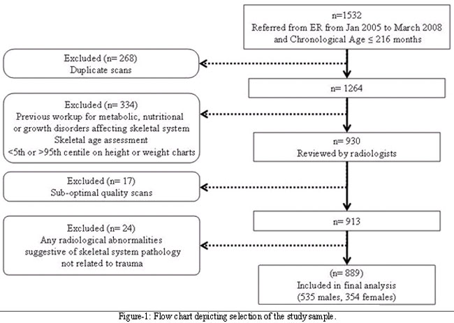
On the basis of patient identity, 268 radiographs were ascertained duplicate and were excluded. Next, the patient charts were reviewed to exclude patients who fulfilled any of the following criteria: a) Previous workup for suspected metabolic, nutritional or growth disorders b) Previous SA assessment c) Below 5th or beyond 95th centile on National Center for Health Statistics reference charts for either height or weight.24 At this stage 334 scans were excluded.
Two paediatric radiologists, each with more than ten years of experience, independently reviewed a total of 930 scans. The observers were blinded to the CA of the subjects. SA was assessed according to the Greulich-Pyle atlas.9 Right hand radiographs were used in case of non-availability of left hand radiographs.3 During review, 17 scans were judged to be of sub-optimal quality whereas radiographic evidence of skeletal system pathology unrelated to trauma was found in 24 scans. These scans were also excluded resulting in a final sample of 889 scans.
Children of each gender were further divided into four groups.2,3 For males, Early Childhood 0-45 months; Middle Childhood 46-90 months; Late Childhood 91-159 months; Adolescence 160-216 months. For females, Early Childhood 0-46 months; Middle Childhood 47-100 months; Late Childhood 101-159 months; Adolescence 160-216 months. Mean CA and SA were calculated for each group and any significant differences between the two were established through two-tailed, paired t-test at a level of significance a < 0.05.
After an interval of four weeks, one hundred randomly selected radiographs were reviewed independently by both reviewers, blinded to the CA at this point too. Pearson\'s Correlation coefficient (r) was calculated to assess inter-observer reliability and paired t-test was used to identify any significant difference between SA estimations by the two observers. SPSS version 16 was used for all statistical analyses.
Results
A total of 889 Hand-Wrist radiographs were included in the final analysis. No significant difference was found between skeletal age assessments by the two observers (Mean difference = 0.4 months, p=0.584). The Pearson\'s correlation coefficient was computed to be 0.992 (p<0.001). The scatterplot is illustrated in Figure-2.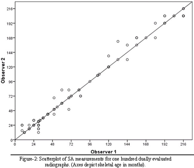
Mean differences of up to 13 months were observed between SA and CA. The differences were statistically significant in all groups except adolescent males. (p<0.05) These findings are elaborated in Table-1.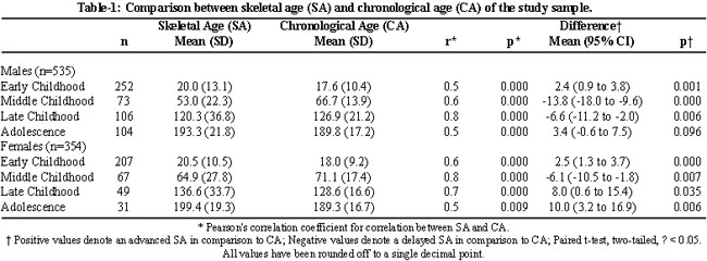
Discussion
We found significant differences between SA assessed by GP atlas and CA in a subset of Pakistani children. In males, SA was advanced during early childhood, delayed during middle and late childhood and, again, advanced during adolescence. In females, the trend was similar except for advanced SA in late childhood. The confidence intervals for the mean difference were wider in the older age groups. This may be a reflection of the individual variability since the inter-observer agreement was excellent in the current study and the same may be presumed about intra-observer reliability in view of the observers\' experience.10,12,13,18
Previous explorations in Larkano, Pakistan also indicated that skeletal maturation of Pakistani children does not conform to the standards of Greulich and Pyle.7,21 In children of Larkano, SA matched CA during initial years of life. This was followed by a delay till CA of 15 years in boys and 13 years in girls, the approximate age of puberty for the respective sex. Subsequently, SA was advanced, with complete maturity being attained at 16 years in females and 18 years in males.7,21 The trends in Larkano and Karachi are almost similar, except for an earlier changeover in females in the present study. This discordance could be attributable to dissimilar categorization of study samples or to genuine variations of skeletal development between the two study populations but calls for further research to elucidate this point.
Conceivably, both \'nature\' and \'nurture\' may play a role in the variation of skeletal maturity across diverse populations. Genetic profile, SES, nutrition and long term well being are some of the factors that have been pointed to in this regard.7,21 Previous explorations suggest that the trends of skeletal maturity may vary across ethnicity within same locale,2,3,12 across geographical regions within genetically akin populations,25 and even across time within the same population in the same country.5,20
Investigations in Turkey documented trends similar to our population, with delayed SA before and advanced SA after puberty, and adjudged GP inapplicable to Turkish children.8,15 In Spanish children, aged 0-14 years, a delay of about three months was observed in males but a good fit to the GP atlas was reported in case of females.16 On the other hand, GP atlas still remains applicable to North European,13 Central European,18 and South Australian children.20 The socio-economic variables differ little within these nations. However, on the genetic spectrum, Turkey is comparatively close to South Asia whereas Spain is the distant cousin in the European lineage.26 At the risk of over-simplification, a parallel may be drawn between the genetic distance and the SA variations, suggesting that \'nature\' may have a large hand in shaping skeletal maturity.
Studies from the other side of the Atlantic, reporting SA variations across diverse ethnicities within the same locale, also point in the same direction.2,3,12 However, quite remarkably, these studies discovered digression from GP atlas even in European-American children.3,12 Likewise, secular increases in skeletal maturation have been identified in Australian males.5,20 On the other hand, GP atlas was found befitting for 12-28 month old, affluent Malay children17 whose genetic make up, social practices and nutrition are distinctly diverse from the GP population. In the same vein, other studies including the present one, have documented the difference between SA and CA to be the least during the initial years of life.2,3,7,12,21 These pieces do not fit perfectly; but they do allude to a place for \'nurture\' in the big picture. Nevertheless, few studies that have actually probed theoretically plausible connection have failed to demonstrate any direct link.14,19 It may be speculated that skeletal maturity is a dynamic parameter influenced by the genetic make up of a population, its SES, environmental milieu and location on the epidemiological curve at a given point in time. Further prospective explorations taking these aspects into account should make the picture more vivid.
There are some limitations of this study that should be considered before drawing any implications from its findings. Firstly, though we made the best of our efforts to select healthy children, variables such as SES and anthropometrical measurements were not controlled in this retrospective comparison. Secondly, established information regarding puberty of Pakistani children is lacking. In addition, there is no study in South Asia that could be used as a precedent. Hence, the grouping of study sample followed the methods of foreign investigators.
Conclusions
The findings of this study suggest against the applicability of GP atlas to Pakistani children. We propose a cautious approach while employing GP atlas in this population in order to ensure appropriate clinical and medico-legal decisions.
Acknowledgements
The authors would like to thank Ms. Aimon Fatima for her invaluable help with the manuscript preparation.
References
1.Cox LA. The biology of bone maturation and aging. Acta Paediatr Suppl 1997; 423: 107-8.
2.Loder R, Estle D, Morrison K, Eggleston D, Fish DN, Greenfield ML, et al. Applicability of the Greulich and Pyle skeletal age standards to black and white children of today. Am J Dis Child 1993; 147: 1329-33.
3.Ontell FK, Ivanovic M, Ablin DS, Barlow TW. Bone age in children of diverse ethnicity. AJR 1996; 167: 1395-8.
4.Zerin JM, Hernandez RJ. Approach to skeletal maturation. Hand Clin 1991; 7: 53-62.
5.Ranjitkar S, Lin NH, Macdonald R, Taylor JA, Townsend GC. Stature and skeletal maturation of two cohorts of Australian children and young adults over the past two decades. Aust Orthod J 2006; 22: 47-58.
6.Yeon KM. Standard bone-age of infants and children in Korea. J Korean Med Sci 1997; 12: 9-16.
7.Rikashor RM, Qureshi AM, Rathi SL, Channa NA. Skeletal maturity in Pakistani children. J Anat 1999; 195: 305-8.
8.Büken B, Safak AA, Yazici B, Buken E, Mayda AS. Is the assessment of bone age by the Greulich-Pyle method reliable at forensic age estimation for Turkish children? Forensic Sci Int 2007; 173: 146-53.
9.Greulich WW, Pyle SI. Radiographic atlas of skeletal development of the hand and wrist. 2nd ed. Stanford: Stanford University Press; 1959.
10.King DG, Steventon DM, O\'Sullivan MP, Cook AM, Hornsby VP, Jefferson IG, et al. Reproducibility of bone ages when performed by radiology registrars: an audit of Tanner and Whitehouse II versus Greulich and Pyle methods. Br J Radiol 1994; 67: 848-51.
11.Oestreich AE. Tanner-Whitehouse versus Greulich- Pyle in bone age determinations. J Pediatr 1997; 131: 5-6.
12.Mora S, Boechat MI, Pietka E, Huang HK, Gilsanz V. Skeletal age determinations in children of European and African descent: applicability of the Greulich and Pyle standards. Pediatr Res 2001; 50: 624-8.
13.van Rijn RR, Lequin MH, Robben SG, Hop WC, van Kuijk C. Is the Greulich and Pyle atlas still valid for Dutch Caucasian children today? Pediatr Radiol 2001; 31: 748-52.
14.Freitas D, Maia J, Beunen G, Lefevre J, Claessens A, Marques A, et al. Skeletal maturity and socio-economic status in Portuguese children and youths: the Madeira growth study. Ann Hum Biol 2004; 31: 408-20.
15.Koc A, Karaoglanoglu M, Erdogan M, Kosecik M, Cesur Y. Assessment of bone ages: is the Greulich-Pyle method sufficient for Turkish boys? Pediatr Int 2001; 43: 662-5.
16.Jiménez-Castellanos J, Carmona A, Catalina-Herrera CJ, Vinuales M. Skeletal maturation of wrist and hand ossification centers in normal Spanish boys and girls: a study using the Greulich-Pyle method. Acta Anat (Basel) 1996; 155: 206-11.
17.Chen ST, Jee FC, Mohamed TB. Bone age of Malaysian children aged 12 to 28 months. J Singapore Paediatr Soc 1990; 32: 97-101.
18.Groell R, Lindbichler F, Riepl T, Gherra L, Roposch A, Fotter R. The reliability of bone age determination in central European children using the Greulich and Pyle method. Br J Radiol 1999; 72: 461-4.
19.Lewis CP, Lavy CB, Harrison WJ. Delay in skeletal maturity in Malawian children. J Bone Joint Surg Br 2002; 84: 732-4.
20.Lin NH, Ranjitkar S, Macdonald R, Hughes T, Taylor JA, Townsend GC. New growth references for assessment of stature and skeletal maturation in Australians. Aust Orthod J 2006; 22: 1-10.
21.Shaikh AH, Rikhasor RM, Qureshi AM. Determination of skeletal age in children aged 8-18 years. J Pak Med Assoc 1998; 48: 104-6.
22.World Health Organization. WHO Statistical Information System. (Online) 2008 (Cited 2009 Jan 5). Available from URL: http://www.who.int/whosis/whostat/2008/en/index.html.
23.City District Government Karachi. Official Web Portal. (Online) 2009 (Cited 2009 Jan 11). Available from URL: http://www.karachicity.gov.pk/.
24.Hamill PVV, Drizd TA, Johnson CL, Reed RB, Roche AF, Moore WM. Physical growth: National Center For Health Statistics percentiles. Am J Clin Nutr 1979; 32: 607-29.
25.Ashizawa K, Kumakura C, Zhou X, Jin F, Cao J. RUS skeletal maturity of children in Beijing. Ann Hum Biol 2005; 32: 316-25.
26.Bauchet M, McEvoy B, Pearson LN, Quillen EE, Sarkisian T, Hovhannesyan K, et al. Measuring European population stratification with microarray genotype data. Am J Hum Genet 2007; 80: 948-56.
Journal of the Pakistan Medical Association has agreed to receive and publish manuscripts in accordance with the principles of the following committees:


