

M. N. Khan ( The Aga Khan University Medical Center. Karachi. )
Y. Bhurgri ( The Aga Khan University Medical Center. Karachi. )
S. Gill ( The Aga Khan University Medical Center. Karachi. )
M. I. Nasir ( The Aga Khan University Medical Center. Karachi. )
T. Siddiqui ( Departments of Pathology and Medicine, The Aga Khan University, Karachi. )
N. Kayani ( The Aga Khan University Medical Center. Karachi. )
March 2003, Volume 53, Issue 3
Original Article

Abstract
Objective:To review cases of male breast cancer.
Settings: Histopathology Section, Department of Pathology, The Aga Khan University, Karachi.
Method: Fifty one cases of male breast cancer specimens, received during a period of 10 years, routinely processed and stained with Haematoxylin and Eosin were analyzed. Special stains and Immunohistochemistry were used in difficult cases.
Results: Male breast cancer affected individuals in the sixth and seventh decades of life with a mean age of 56.2 years. Infiltrating ductal carcinoma (IDC) was the predominant type. Skin involvement was seen in 27.45% of the cases. Breast lump was the most common presenting symptom followed by skin ulceration. At the time of presentation 43.13% patients had a tumour size of more than 3 cm.
Conclusion: Male breast cancer is a rare disease. Most of our findings correspond to the published local and international data (JPMA 53:114;2003).
Introduction
Male breast cancer is a rare disease, it accounts for less than 1% of all cancers and 0.1% of all deaths from cancer in men.1 Breast cancer is 100 times less common in men as compared to women and affects predominantly older populations. with a peak incidence at approximately 60 years of age.1,2 Ductal carcinoma is the predominant type, which presents at a more advanced stage and seems to carry a less favorable prognosis than in its female counterpart. However a poor prognosis is not fully substantial for every study when stage matched outcomes are taken into account,1,3-6 Most of the patients present with painless firm breast lumps)1,7.8 Some of the studies have reported nipple discharge as a presenting symptom.9,10 The other signs and symptoms include nipple retraction, skin ulceration, skin erythema, breast tenderness, gynaecomastia etc. Tumour size and axillary lymph node status are considered as important prognostic factors.1 A prevalence of male breast cancer and its distribution has been examined in a number of studies in the West, however, such studies are almost non-existent for the Pakistani population. In this study we have analyzed the data of male breast cancer diagnosed from July 1991 to June 2001, with a view to have preliminary information about this tumour.
Material and Methods
This was a retrospective study conducted in the histopathology section, department of Pathology, the Aga Khan University Hospital, between July 1991 to June 2001. It includes only the surgical specimens and cytology fluids were not studied. The specimens were grossed according to the standard procedure. In the lunipectomy specimens two sections were taken from the lesion and at least one section from the margin of excision. The tumor size and the gross findings were noted. In simple mastectomy specimens additional sections were taken from the skin, nipple and areola. At least one section was taken from each recovered lymph node, where available. The specimens were routinely processed as these were received fixed in 10% formalin. After processing, 4-5m sections were cut and stained with Haematoxylin and Eosin stains. Special stains such as Perioidic Acid Schiff, and Reticulin were used when required. Immunohistochemistry (Peroxidase and antiperoxidase techniques) was used in difficult cases. The markers included Cytokeratin MNF. Cytokeratin CAM 5.2, Cytokeratin AEI/AE3. LCA. Vimentin. Desmin and Actin.
Results
During the 10 year period of the study, a total of 2,13,377 surgical specimens were received out of which 53,012 specimens were breast cancers. Of these 53,012 breast cancer cases, only 51(0.096%) were of males, whilst the remaining 52,961 (99.90%) cases were from females. Excision biopsy specimens comprised 26 (50.98%) and mastectomy specimens 22 (43.13%). Needle biopsy was performed in the remaining 3 (5.88%) cases.
Right breast involvement by the tumor was seen in 21 (41.76%) and left breast in 15 (29.41%) cases. In I (1.96%) case, bilateral involvement was found. In 14 (27.45%) specimens the side of involvement was not known.
Most of our patients at the time of diagnosis were in the sixth and seventh decades of life. (Figure).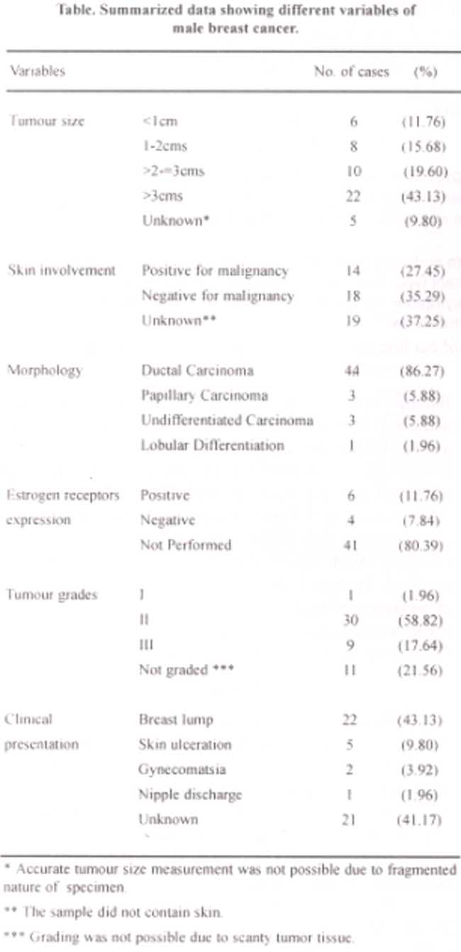
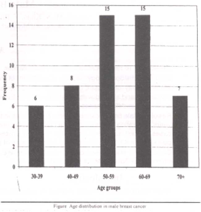
The ages ranged from 33 to 82 years with a mean age of 56.2 years and median of 55 years.
Out of 51 cases a clinical history was provided in 30 of whom 22 (43.13%) presented with a breast lump. Five patients (9.80%) presented with skin ulceration, two (3.92%) with gynecomastia and one (1.96%) with nipple discharge. In 22 (43.13%) cases the tumour size was greater than 3 cms, in 10 (19.60%)>2-3 ems, 8 (15.68%) between 1-2 ems and in 6 (11.76%) cases, the tumour was less than 1 cm in size. In 5 (9.80%) the exact tumour size was not known due to the fragmented nature of the biopsy.
Inter ductal carcinoma (IDC) was the predominant diagnosis in 44 (86.27%) cases,followed by papillary carcinoma in 3 (5.88%). Lobular differentiation was seen in 1(1.96%) and in 3 (5.88%) specimens. the tumor was undifferentiated. Modified Bloom Richardson system was used for tumour grading. One (l.960/o) was grade 1, 30 (58.82%) were grade II and 9(17.64%) were grade III. In 11 (21.56%) specimens grading was not done due to the small size of the biopsy material.
Skin involvement was found positive in 14 (27.45%) cases which included 4 (7.8%) with a pagetoid spread. Skin involvement was negative in 18 (35.29%) cases. In the remaining 19 (37.25%) skin was not present to assess invasion.
Estrogen receptor status was studied in only 10 (19.60%) patients. Of these 6(1 1.76%) were positive and 4 (7.84%) cases negative. In the remaining 41 (80.39%) estrogen receptor studies were not performed.
Discussion
In our analysis the incidence of male breast cancer represents 0.09% of all breast cancers. In most other regional and international studies it was found to be less than 1%)1.5 In an extensive study conducted in over 100 cancer registries all over the world, the incidence of male breast cancer was found to be 0.5/100,000 annually.8 Lower figures of male breast cancer may be due to the fact that males have lower breast mass as compared to females. Secondly as compared to fully functional female breast, it is a vestigial organ in the male. On the contrary in some areas of Africa the prevalence of male breast cancer is found to be as high as 6-l5% of all male carcinomas.8 Unlike female breast cancer almost all the studies on male breast cancel are retrospective due to rarity of the disease.5
As observed in other studies, breast lump was the most common presenting symptom in the presented analysis.1,5 Breast lump is the initial symptom noted in both sexes regardless of the fat content of the breast. However the superficial nature of the male breast makes it easier to detect. Some studies have reported nipple discharge as a presenting symptom of male breast cancers and suggest that it should always be investigated by biopsy.9.10 In our study only one patient complained of nipple discharge while five presented with other skin complaints like ulceration.
In this study most patients (59%) were in the sixth and seventh decades of life. International other reports have shown male breast cancer to affect older individuals.1,5,11,12 In comparative studies the mean age of breast cancer patients was found to be approximately ten years older in comparison to the females.11.12 Exact cause for this is not known. The impact of hormonal influences on the pathogenesis of breast cancer in the males, remains undetermined. In our study the mean age at the time of diagnosis was 56.2 years, which is somewhat less then other international studies. However it is still higher than the mean age of female breast cancer.
In 43.13% cases tumor size was found equal to or greater than 3 ems at the time of diagnosis. Considering the superficial location and low amount of fat in male breast, it should be considered a diagnostic delay. This also draws our attention to the flaw in our health care system to detect such obvious lesions so late.
In this study IDC was the most common diagnosis. Literature search revealed almost all the IDC to be the most common malignancy in men1.7.8.13 In our study there was only a single case of breast carcinoma showing lobular differentiation. In males due to the absence of lobules, lobular carcinoma is almost a non-existent entity.8,13 In a case reported by Michaels BM, et al (1994), a single case of lobular carcinoma was found in a genotypically proven male.14 However in our study the karyotype was not established.
Due to low breast fat and close proximity with skin; the involvement of the latter is common in males as compared to females.7,15 Xu et al observed skin lesions in half their cases)16 In this study skin involvement was seen in 14 (27.45%) of the total cases. In a regional study at Armed Forces Institute of Pathology (AFIP) Rawalpindi Pakistan. skin involvement was seen in 36% of the cases, which is some what higher than our findings.7
We performed Estrogen receptor analysis in 10 cases of which 6 were positive. Multiple studies from the western hemisphere showed similar result.1,17 A study from Norway showed Estrogen receptor positivity in 72% cases.17 Estrogen receptor status is important while considering tamoxifen as adjuvant therapy.
This study is one of the few initial studies on the subject in our part of the world. With better collaboration between clinicians and pathologists and the introduction of molecular technology, more advanced and comprehensive studies are expected in the future.
References
1.Memon MA, Donohue JH. Male breast cancer. Br J Surg 997:84: 433-35.
2.Gibson TN. Brady-West D, Williams E, et al. Male breast cancer. An analysis of four cases and review ofthe literature. W Indian Med.J 2001;50:165-8.
3.Gough D. Donahue JH. Evans MM. et al. A 50-year experience of male breast cancer. is outcome changing? Surg Oncol 1993:2:325-33
4.Wagner JL. Thomas CR Jr, Koh WJ, et al. Carcinoma of the male breast. update 1994. Med Pediatr Oncol 1995:24:123-32.
5.Willsher PC. Leach IH. Ellis 10. et al. A comparison outcome of male breast cancer. Am J Surg 997. 173185-8
6.Jepson AS, Fentiman IS. Male breast cancer. Int J Clin Pract 998:52.571-6
7.Jamal S. Mushtaq S. Malik IA, et al. Malignant tumours of the male breast a review of 50 cases. J Pak Med Assoc 1994: 44:275-7.
8.Nagadowska MM. Fentiman IS. Male breast cancer. Br J Hosp Med 1993:49:104-6.
9.Spence R, MacKenzie G, Anderson J, et al. Long term survival following cancer of male breast in Northren Ireland. Cancer 1985:55:648-52.
10.Treves N. Robbins G, Amoroso W. Serous and serosanguineous discharge from the male nipple. Arch Surg 1956:73:319-29.
11.Borgen P. Wong GY, Vlamis V. et al. Current management of male breast cancer Ann Surg 1992. 215:451-8.
12.Riberio G. Male breast carcinoma. a review of 301 cases from Christie Hospital and Holt Radium Institute. Manchester. Br J Cancer 1985;51:11 5-19
13.Willsher PC. Leach lH, Ellis 10, et al. Male breast cancer pathological and immunohistochemieai features. Anticancer Res 1997; 17:2335-8.
14.Michaels BM, Nunn CR, Roses DF. Lobular Carcinoma of the male breast. Surgery 1994:115:402-5.
15.Norris HJ, Taylor HB. Carcinoma ofthe male breast. Cancer 1969:23:1428-35
16.Xu RN. Male breast cancer: clinical, pathologic and immunohistocheinical study. Zhonghua Wai Ke Za Lhi 989:27.28-30.
17.Andre S. Fonseca I, Pinto AE, et al. Male breast cancer a reappraisal of’ clinical and biologic indicators of prognosis. Acta Oncol 2001:40:472-8.
Journal of the Pakistan Medical Association has agreed to receive and publish manuscripts in accordance with the principles of the following committees:


