

Maryam Sohrabi ( Department of Anatomical Sciences, Faculty of Medicine,Tabriz University of Medical Sciences, Tabriz, Iran. )
Jafar Soleimani, ( Department of Anatomical Sciences, Faculty of Medicine,Tabriz University of Medical Sciences, Tabriz, Iran. )
Leila Roshangar ( Department of Anatomical Sciences, Faculty of Medicine,Tabriz University of Medical Sciences, Tabriz, Iran. )
Seda Vatansever ( Department of Histology - Embryology, Faculty of Medicine, Celal Bayar University of Medical Sciences. Manisa, Turkey )
Fatemeh Arbabi ( Department of Oral Disease, Faculty of Dentistry, Zahedan University of Medical Sciences, Zahedan, Iran. )
Amir Afshin Khaki ( Department of Anatomical Sciences, Faculty of Medicine,Tabriz University of Medical Sciences, Tabriz, Iran. )
Mehran Mesgari Abbasi ( Drug Applied Research Center,Tabriz University of Medical Sciences, Tabriz, Iran. )
Yosuf Dustar ( Department of Veterinary, Pathology College of Veterinary Medicine, Islamic Azad University (Branch of Tabriz University), Tabriz-Iran. )
Yosuf Javadzadeh ( Department of Pharmaceuitics, Faculty of Pharmacology, Tabriz University of Medical Sciences, Tabriz, Iran. )
November 2009, Volume 59, Issue 11
Original Article

Abstract
Objective: To compare the preventive effect of dietary and topical Celecoxib (CCB), a potent inhibitor of COX-2 on 4-NQO-induced tongue SCC in rat.
Methods: Fifty male Sprague Dawley adult 3- 3.5 months old rats were used as animal model in this study. The tongue SCC was induced by a daily administration of 30 ppm 4-NQO, in drinking water, for 8 months. The rats in case groups received dietary or topical CCB. Tongue Specimens were prepared for histopathological and immunohistochemical (Ki-67) staining and or TUNEL assay were examined. Values are expressed as mean ± SEM and analyzed with Npar Kruscal Wallis and one-way ANOVA tests. p < 0.05 was considered significant.
Results: The incidence of tongue precancer lesions, judged by morphological and morhometrical criteria and apoptosis/proliferation ratio, was significantly (p<0.01) reduced by CCB. The effect of topical CCB use, at high doses, was comparable to the effect of dietary CCB.
Conclusion: Both topical and dietary CCB have inhibitory effect on 4-NQO induced SCC on tongue. The effect of CCB is probably mediated by suppression of cell proliferation and induction of apoptosis (JPMA 59:769; 2009).
Introduction
The incidence of oral cancer, the major site being the tongue, exhibits marked geographic variation, with the highest morbidity and mortality rates appearing in southern Asia, where people have the habit of chewing betel quid and tobacco, but recently it has been increasing worldwide, particularly in young adults.1 Despite recent advances in surgical procedures, radiotherapy and chemotherapy, oral cancer remains a major problem. One reason is the characteristic "field cancerization," with relatively high frequencies of second primary cancers at different sites.2
(Cycloxygenases), COXs, a rate-limiting enzymes for producing prostanoids, consist of two isomers, COX-1 and COX -2. COX-2, is an inducible immediate early gene that has recently been postulated to be involved not only in inflammation but also in carcinogenesis, with impact on cell proliferation, differentiation, apoptosis, angiogenesis, metastasis, and immunological surveillance.3 COX-2 overexpression leads to an increased production of prostaglandins, including prostaglandin E2, which stimulates epithelial cell proliferation and inhibits apoptosis.2
Administration of 4-NQO, in drinking water, can induce tumours in oral cavities in mice and rats.4 Rat oral Squamouse Cell Carcinomas induced by 4-NQO shows morphological and histopathological similarities to those of human tumours, extensively been used to investigate and test a wide variety of synthetic and natural agents for their chemopreventive potentials.5
A variety of Non Steroidal Anti Inflammatory Drugs (NSAIDs) were shown to be capable of stimulating apoptosis.6 Therfore in current years, many similar agents such as celecoxib were tested and found to be effective chemoprotectors. COXs have been postulated to be a target for NSAIDs and selective COX-2 inhibitors, prevent the formation and growth of tumours in experimental animals.
Celecoxib, a potent cyclooxygenase 2 (COX-2) inhibitor is currently being used as an anti-inflammatory agent for the treatment of rheumatoid arthritis and osteoarthritis.7 CCB also is under investigation for the treatment of various malignant and premalignant tumours, including colorectal, breast, lung and prostate cancer.8
However, long-term oral administration of high doses of Cox-2 inhibitors such as celecoxib may lead to systemic toxicity.9 The aim of this study is to examine whether the effect of topical CCB is comparable to dietary CCB and using topical CCB is a suitable substitute of dietary CCB could be substituted by topical CCB with the same chemopreventive effect for tongue carcinoma?
Materials and Methods
Animals: A total of 50 adult male Sprague Dawley rats (3-3.5 month old) were used in the experiment. All rats were obtained from Tabriz Medical University animal-house and at first quarantined and acclimatized to laboratory conditions for 2 weeks. During the experiment, each rat was housed, in a metal cage, with hardwood chips for bedding, in an air-conditioned room with a 12-h light/12-h dark cycle.
Chemicals: 4-NQO was purchased from Sigma Inc. (Germany) and CCB from Aldrich Inc. (Germany).
4-NQO solution (30 ppm) was prepared thrice a week by dissolving the carcinogen in distilled water and was given in light-opaque covered bottles. Rats were allowed access to the drinking water at all times during the treatment except 2-3 hours after adhesive gels usage.
Diets containing 2000 ppm CCB were prepared thrice a week. CCB powder was added to wet powdered basal diet and then dried in oven (50°C). Each rat received 2000 ppm CCB with daily food.
Adhesive Gels were prepared by mixing the Base Plasty with Oral Paste including 2000 ppm or 3000 ppm CCB.
For Base Plasty preparation: liquid paraffin and LDPE (Low Density Polyethylene) were mixed with 9.5 to 0.5 proportion.
For Oral Paste preparation: pectin,gelatine and sodium carboxy methyl cellulose 1/1/1 were mingled.
Then Oral paste was added to 2000 ppm or 3000 ppm CCB, mixed with Base Plasty precisely. These adhesive gels were put in plastic containers into 4oC until using, when they were brought to room temperature.
Experimental Procedure: The rats were randomly assigned to one of five treatment group. These groups include: group 1) 2000 ppm dietary CCB as control; group 2) 30 ppm 4-NQO treatment; group 3) 30 ppm 4-NQO+2000 PPM dietary CCB treatment; group 4) 30 ppm 4-NQO+2000 ppm topical CCB treatment; group 5) 30 ppm 4-NQO+3000 ppm topical CCB treatment. All animals procedures complied with an approved TUMS animal care protocol and the Tabriz university of medical science animal care and use committee.
Body weights, water and food consumption of animals were measured thrice a week. The tongues was checked regularly fifth time during study for disorders including: leukoplakia, erytroplakia, erytroleukoplakia, and tumour existance.
At the end of the 8 weeks, animals were killed under ether anaesthesia. Their whole tongues were incised and then oral part (2/3 anterior) longitudinally cut into two slices and each of them again cut into two halves. Specimens after fixation in 10% phosphate-buffered formalin for 48 hours prepared for light microscope, routinely embedded in paraffin, and was sectioned serially. The serial 5 µm sections were stained with H&E, Ki-67 and TUNEL staining.
For Ki-67 staining, Cell Proliferation Detection Kit was utilized and staining with Ki-67 antigen monoclonal mouse anti-rat (code No.7248) was carried out according to factory instruction. The Ki-67 positive cells appeared brown. For TUNEL stainig In Situ Cell Death Detection Kit (Cat. No.11 684 817 910) was purchased from Roche-Germany The TUNEL positive cells appeared brown.
Evaluation of Sections
Specimens were qualified blindly by one histologist and one pathologist. Histological slides (H&E) were evaluated by using a light microscope (Olympus BX40, Tokyo, Japan). In the H&E stained slides, thickness of tongues epithelia at five different fields were measured by software Motic-image plus 2 and mean of total number considered as thickness of epithelium layer. Morphological study of epithelium in the specimens including basal layer disorder, dividing cells in basal and prickle layers, size of the prickle cells, multilayered basal cells and rounded basal cells were scaled as (0=None 1=Low 2=High 3=Very high). Pathological changes including dysplasia, hyperplasia, hyperkeratosis, parakeratosis, tumour like cell and pearl body existence were determined in the epithelia and were scaled on the basis of WHO criteria10 as Intact, Mild, Moderate and Severe.
Histochemical slides (TUNEL) were evaluated by using a light microscope (Olympus BX40, Tokyo, Japan) and a fluorosence microscope (Olympus IX71, Tokyo, Japan).
Average number of epithelial apoptotic cells was estimated by two observers blinded to the experimental conditions through counting TUNEL positive cells in five randomly medium-power fields (400X). The percentage of positive cells with TUNEL assay served as rate of apoptosis. Cells in areas with necrosis, poor morphology or in the margins of sections were discarded. Immunohistochemical slides (for Ki-67), were evaluated by using a light microscope (Olympus BX40, Tokyo, Japan). Average number of epithelial proliferative cells was estimated through counting positive cells in five random neighbouring medium-power fields (400X) that included 100 cells (at least 500 cells in each lesion) and dividing the total to five.
Statistical Analysis
The difference between means±SE for control and experimental groups were examined by using one-way ANOVA and Npar Kruscal Wallis tests. Statistical difference of p-value at the level of 0.05 or less was considered significant.
Results
Animal Body Weight and Water and Food Intake: Animal weight determinations are shown in Figure-1. As the figure shows at the end of the experimental period, the mean final body weight in all groups, in comparision to control group,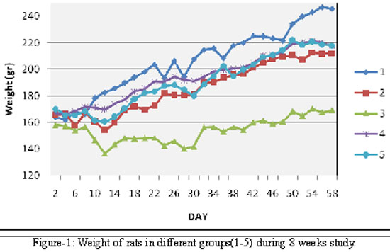
decreased significantly (p=0.001).
There were significant differences (p<0.0005) between average Intake of drinking water containing of 4-NQO in and Intake of food with or without CCB, in different groups with control as shown in Table-1.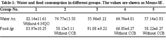

Macroscopic Changes: The dorsal aspect of the tongue was examined for macroscopic changes. In the control group there was not a considerable change in the epithelium, in group 2, which received 30 ppm 4-NQO, epithelium showed small whitish leukoplakial, reddish erythroplakial and protuberant tumour lesions, varying in size from 1 mm to 1.5 mm in diameter. The number and size of such lesions tend to decrease in CCB treated groups (groups 3-5), especially in group 5 (received 3000 ppm CCB as topical gel).
Epithelial Thickness: Determination of thickness of lingual epithelium by Motic Image-Plus 2 revealed that mean of epithelial thicknesses (without keratin layer) were similar in all groups and the difference between groups were not signigicant (p=0.60).
Morphological Studies: Microscopy revealed that in comparison to control group: frequency of large prickle cells were significantly (p=0.01) higher in experimental groups. Occurence of multilayered basal cells according to their scale were lower in groups 4,5 (topically received CCB) in comparison to group 3 (dietary CCB). Number of round basal cells was higher in groups 2 and 5 (p=0.00) in comparison to other groups.
Histopathological Studies: Microscopy revealed that different histopathological changes such as dyskeratosis, hyperkeratosis, parakeratosis, dysplasia, tumour like cells and pearl bodies appeared in treated groups (Figure 3a 1-5).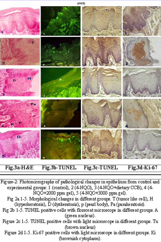
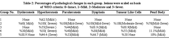
These changes were maximum in group 2 (Figure 3a 2-5) and decreased by CCB treatment (Figure 3a 2-5). The changes were quantified as determining the percentages of affected cells and scaled as mild-severe according to their characteristics (Table-2).
Immunohistochemical Studies: TUNEL positive cells appeared brown with light microscope (Figure 2c 1-5) and green fluorescent with fluorescent microscope (Figure 2b 1-5). Ki-67 positive cells appeared goldish-brown with light microscope (Figure 2d 1-5) The TUNEL and Ki-67 positive cells were randomly counted in 5 different fields and apoptotic or proliferative indexes were calculated. Then apoptosis/proliferation ratio, in each group, was calculated and compared with other groups. As shown in Table-3, the ratio was least in group 2 and increased by CCB treated with maximum value in group 3.
Other observation:
1- During the study four animals died (three from group 2 and one from group 4). Survival rate were 95.8%, 97.9%, 97.9% at days 17, 18, 19.
2- After animal dissection, lungs, kidneys and livers were larger than normal in groups 2 and 4 but in groups (3 and 5) they had moderate size.
3- There was a weak gasterointestinal tract bleeding only in group 3 and 4.
Discussion
The aim of the present study was to induce tongue cancer by 4-NQO and then evaluate and compare, chemopreventive effect of dietary and topical CCB treatment. Our results showed that 4-NQO induced typical precancerous signs and morphological alterations on the tongue epithelium. These changes included weight loss, macroscopic lesions, changes in cell size and pathological changes. In accordance with our findings it is shown, that 4-NQO produces SCCs in mice and rats.4 4-NQO is a water soluble quinoline derivative that can cause DNA adduct formation and also can undergo redox cycling to produce reactive oxygen species that result in mutations and DNA strand breaks.11
Oral leukoplakia is the most common premalignant lesion of oral cancer, and up to 20% of the patients with leukoplakia develop invasive carcinoma.12 Oral cancer usually develops from hyperplasia through dysplasia to carcinoma in the manner of `field cancerization' due to carcinogen exposure.13 Dysplasia was characterized by increased number of basal cells, irregular epithelial stratification, increased number of mitotic figures, increased nuclear-to-cytoplastic ratio and loss of polarity of basal cells. According to the field cancerization concept, key molecular and biochemical events are thought to occur before altered cellular morphology is apparent.
Judging from TUNEL and Ki-67 assays revealed that proliferation was high, apoptosis was low and apoptosis /proliferation was minimum in 4-NQO treated group. There was a high induction of apoptosis and low expression of Ki-67 with CCB treatment. The apoptosis/proliferation ratio was maximum on dietary CCB and it was similar to high doses of topical CCB treatment. The result indicate that while topical CCB treatment is effective for chemoprevention but higher doses should be used in comparison to dietary CCB treatment.
Apoptosis is a genetically regulated active process in which changes in cellular architecture occur, resulting in self-destruction of the cell. Apoptosis is complementary to mitosis in regulating cell populations in both physiological and pathological conditions.14 Cell proliferation is regarded as one of the most important biological mechanisms in oncogenesis.15 Cell proliferation in tissues of experimental animals is essential for toxicology and carcinogenesis studies and also to assess the efficiency of cytotoxic and chemopreventive drugs in cancer research.
Cox2 has been known to play an important role in the initiation and post-initiation stages of carcinogenesis. COX-2 mRNA and protein expression is inducible in most tissues by external stimuli such as tumour promoters, growth factors, and cytokines. Cox2 was barely detectable in the normal epithelium but up-regulated in hyperplasia and further overexpressed in dysplasia and squamous cell carcinoma. Up-regulation of COX-2, is associated with a variety of carcinogenic mechanisms. These mechanisms include abnormal expression of epithelial growth factors, epithelial and microvascular proliferation, resistance to apoptosis, enhancing angiogenesis and suppression of antitumour immunity.16
In human oral cancer, overexpression of Cox2 has been well documented.16 Direct evidence of COX in tongue carcinogenesis has been shown in a rat model with inhibition of dysplasia occurring with the administration of NSAIDs in animal models. Another study revealed that the administration of an NSAID reduced the incidence of squamous cell carcinoma in animals to 23-31% compared with 71% in untreated controls.17 These studies reveal that COX inhibition, particularly COX-2 inhibition, has a potential role in the chemoprevention of head and neck squamous cell carcinomas.
Celecoxib was the first cyclooxygenase 2-selective NSAID approved for the treatment of adult arthritis. CCB exerts potent chemopreventive activity in chemical carcinogen-induced colon, bladder, and breast carcinogenesis and UV-induced skin carcinogenesis also effectively inhibits the growth of colon and breast cancer xenograft tumours in nude mice.18 Currently, CCB is being tested in clinical trials for its chemopreventive or therapeutic activity against various cancers also it has been found to have antitumour activity in tumour cells and tissues that lack the cyclooxygenase 2 enzyme.19 CCB induces apoptosis in various cell types,2- and this activity may account for its chemopreventive and therapeutic activity. However, the mechanisms by which celecoxib induces apoptosis remain largely uncharacterized, Using the DMBA-induced hamster cheek pouch model, Shiotani showed that celecoxib prevented oral carcinogenesis in a dose-dependent manner.21 The reduced proliferation with chemotherapy may be at least partly due to increased apoptosis in actively proliferating cells.22 Daidone et al.further combined the proliferative and apoptotic parameters, which appeared to have an even stronger prognostic value. There are, however, also contradictory data on the role of apoptosis in the prognosis of SCC of the head and neck. Jäckel found that the frequency of apoptotic cells significantly correlated with high mitotic activity, a high malignancy grade of the tumour and disease progression in laryngeal carcinomas.23 This indicates that extensive spontaneous apoptosis balances the high rate of mitosis and thus reflects the growth rate of the tumour. Recently, Celecoxib has been reported to decrease Ki-67 expression in cervical cancer.24 Suppression of cell proliferation as measured by Ki-67 LI has also been reported to accompany polyp regression in a chemoprevention trial in patients with familial adenomatous polyposis (FAP).25 Mao et al. findings provide evidence to support that Celecoxib may be capable of decreasing Ki-67 expression and proliferative activity in bronchial mucosa in continuing smokers.26
Conclusion
The implication suggests that the balance between proliferation and apoptosis rather than their absolute levels could be important and the results suggested that Celecoxib had inhibitory effects against tongue carcinogenesis at the initiation and post-initiation stage and such inhibition may be related to the suppression of cell proliferation, induction of apoptosis. it seems that efficacy of topical CCB is dose dependent. It is an important observation that significant difference was observed between dietary and topical treatments of CCB and topical CCB (3000 ppm) usage is comparable to dietary CCB but If we have seen obviously decreasing animal body weight at dietary CCB (2000 ppm) may simply reflect variations within statistical uncertainty because of the relatively small numbers of animals or because of interaction between 4-NQO and CCB and warrants further studies.
Acknowledgments
The authors are obliged to thank Drug Applied Research Center of Tabriz University of Medical Sciences for their financial support and member of department of Histology — Embryology, Faculty of Medicine, Celal Bayar University of Medical Sciences. Manisa, Turkey for their excellent IHC technical assistance.
References
1.Moore SR, Johnson NW, Pierce AM, Wilson DF. The epidemiology of tongue cancer: a review of global incidence. Oral Dis 2000; 6: 75-84.
2.Turini ME, DuBois RN. Cyclooxygenase-2: a therapeutic target. Annu Rev Med 2002; 53: 35-57.
3.Tsujii M, DuBois RN. Alterations in cellular adhesion and apoptosis in epithelial cells overexpressing prostaglandin endoperoxide synthase 2. Cell 1995; 83: 493-501.
4.Tanaka T, Kojima T, Okumura A, Yoshimi N, Mori H. Alterations of the nucleolar organizer regions during 4-nitroquinoline 1-oxide-induced tongue carcinogenesis in rats. Carcinogenesis 1991; 12: 329-33.
5.Tanaka T. Chemoprevention of oral carcinogenesis. Eur J Cancer B Oral Oncol 1995; 31B: 3-15.
6.Tsutsumi S, Gotoh T, Tomisato W, Mima S, Hoshino T, Hwang HJ, et al. Endoplasmic reticulum stress response is involved in nonsteroidal anti-inflammatory drug-induced apoptosis. Cell Death Diff 2004; 11: 1009-16.
7.Lane NE. Pain management in osteoarthritis: the role of COX-2 inhibitors. J Rheumatol Suppl 1997; 49: 20-4.
8.Patel M, Subbaramaiah K, Du B, Chang M, Yang P, Newman RA, et al. Celecoxib inhibits prostate cancer growth: evidence of a cyclooxygenase-2-independent mechanism. Clin Cancer Res 2005; 11: 1999-2007.
9.Kammerl MC, Debler J, Riegger GA, Kramer BK. COX-2 inhibitors and risk of heart failure. Lancet 2004; 364: 1486-7.
10.Pindborg JJ, Reichart PA, Smith CJ, van der Wall I (eds). World Health Organization. In: Histological Typing of Cancer and Precancer of the Oral Mucosa. 2nd ed. Berlin: Springer-Verlag, 1997; pp 21-31.
11.Phillips DH. Smoking-related DNA and protein adducts in human tissues. Carcinogenesis 2002; 23: 1979-2004.
12.Minter HA, Eveson JW, Huntley S, Elde DJ. Hague A. The cyclooxygenase 2-selective inhibitor NS398 inhibits proliferation of oral carcinoma cell lines by mechanisms dependent and independent of reduced prostaglandin E2 synthesis. Clin Cancer Res 2003; 9: 1885-97.
13.Sudbo J, Ristimaki A, Sondresen JE Kildal W, Boysen M, Koppang HS, et al. Cyclooxygenase-2 (COX-2) expression in high-risk premalignant oral lesions. Oral Oncol 2003; 39: 497-505.
14.Kerr JF, Wyllie AH, Currie AR. Apoptosis: a basic biological phenomenon with wideranging implications in tissue kinetics. Br J Cancer 1972; 26: 239-57.
15.Van Diest PJ, Brugal G, Baak JPA. Proliferation markers in tumours: interpretation and clinical value. J Clin Pathol 1998; 51: 716-24.
16.Wiese FW, Thompson PA, Kadlubar FF. Carcinogen substrate specificity of human Cox-1 and Cox-2. Carcinogenesis 2001; 21: 5-10.
17.Tanaka T, Nishikawa A, Mori Y, Morishita Y, Mori H. Inhibitory effects of non-steroidal anti-inflammatory drugs, piroxicam and indomethacin on 4-nitroquinoline 1-oxide-induced tongue carcinogenesis in male ACI/N rats. Cancer Lett 1989; 48: 177-82.
18.Blumenthal RD, Waskewich C, Goldenberg DM, Lew W, Flefleh C, Burton J. Chronotherapy and chronotoxicity of the cyclooxygenase-2 inhibitor, celecoxib, in athymic mice bearing human breast cancer xenografts. Clin Cancer Res 2001; 7: 3178-85.
19.Hsu AL, Ching TT, Wang DS, Song X, Rangnekar VM, Chen CS. The cyclooxygenase-2 inhibitor celecoxib induces apoptosis by blocking Akt activation in human prostate cancer cells independently of Bcl-2. J Biol Chem 2000; 275: 11397-403.
20.Hashitani S, Urade M, Nishimura N, Maeda T, Takaoka K, Noguchi K. Apoptosis induction and enhancement of cytotoxicity of anticancer drugs by celecoxib, a selective cyclooxygenase-2 inhibitor, in human head and neck carcinoma cell lines. Int J Oncol 2003; 23: 665-72.
21.Shiotani H, Denda A, Yamamoto K, Kitayama W, Endoh T, Sasaki Y, et al. Increased expression of cyclooxygenase-2 protein in 4-nitroquinoline-1-oxide-induced rat tongue carcinomas and chemopreventive efficacy of a specific inhibitor, nimesulide. Cancer Res 2001; 61:1451-6.
22.Daidone MG, Silvestrini R, Luisi A, Mastere M, Benini E, Veneroni S, et al. Changes in biological markers after primary chemotherapy for breast cancers. Int J Cancer 1995; 61: 301-5.
23.Jackel MC, Dorudian MA, Marx D, Brinck U, Schauer A, Steiner W. Spontaneous apoptosis in laryngeal squamous cell carcinoma is independent of bcl-2 and bax protein expression. Cancer 1999; 85: 591-9.
24.Ferrandina G, Ranelletti FO, Legge F, Luriola L, Salutari V, Gessi M, et al. Celecoxib modulates the expression of cyclooxygenase-2, Ki67, apoptosis-related marker, and microvessel density in human cervical cancer: a pilot study. Clin Cancer Res 2003; 9: 4324-31.
25.Sinicrope FA, Half E, Morris JS, Lynch PM, Morrow JD, Levin B, et al. Cell proliferation and apoptotic indices predict adenoma regression in a placebo-controlled trial of celecoxib in familial adenomatous polyposis patients. Cancer Epidemiol Biomarkers Prev 2004; 13: 920-7.
26.Mao JT, Fishbein MC, Adams B, Roth MD, Goodglick L, Hong L, el al. Celecoxib Decreases Ki-67 proliferative index in active smokers. Clin Cancer Res 2006; 12: 314-20.
Journal of the Pakistan Medical Association has agreed to receive and publish manuscripts in accordance with the principles of the following committees:


